3
views
0
recommends
+1 Recommend
0 collections
    0
    shares
      • Record: found
      • Abstract: found
      • Article: found
      Is Open Access

      A New Approach of Fatigue Classification Based on Data of Tongue and Pulse With Machine Learning

      research-article

      Read this article at

      Bookmark
          There is no author summary for this article yet. Authors can add summaries to their articles on ScienceOpen to make them more accessible to a non-specialist audience.

          Abstract

          Background

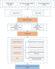
          Fatigue is a common and subjective symptom, which is associated with many diseases and suboptimal health status. A reliable and evidence-based approach is lacking to distinguish disease fatigue and non-disease fatigue. This study aimed to establish a method for early differential diagnosis of fatigue, which can be used to distinguish disease fatigue from non-disease fatigue, and to investigate the feasibility of characterizing fatigue states in a view of tongue and pulse data analysis.

          Methods

          Tongue and Face Diagnosis Analysis-1 (TFDA-1) instrument and Pulse Diagnosis Analysis-1 (PDA-1) instrument were used to collect tongue and pulse data. Four machine learning models were used to perform classification experiments of disease fatigue vs. non-disease fatigue.

          Results

          The results showed that all the four classifiers over “Tongue & Pulse” joint data showed better performances than those only over tongue data or only over pulse data. The model accuracy rates based on logistic regression, support vector machine, random forest, and neural network were (85.51 ± 1.87)%, (83.78 ± 4.39)%, (83.27 ± 3.48)% and (85.82 ± 3.01)%, and with Area Under Curve estimates of 0.9160 ± 0.0136, 0.9106 ± 0.0365, 0.8959 ± 0.0254 and 0.9239 ± 0.0174, respectively.

          Conclusion

          This study proposed and validated an innovative, non-invasive differential diagnosis approach. Results suggest that it is feasible to characterize disease fatigue and non-disease fatigue by using objective tongue data and pulse data.

          Related collections

          Most cited references49

          • Record: found
          • Abstract: found
          • Article: found
          Is Open Access

          Deep Convolutional Neural Networks for Computer-Aided Detection: CNN Architectures, Dataset Characteristics and Transfer Learning

          Remarkable progress has been made in image recognition, primarily due to the availability of large-scale annotated datasets and deep convolutional neural networks (CNNs). CNNs enable learning data-driven, highly representative, hierarchical image features from sufficient training data. However, obtaining datasets as comprehensively annotated as ImageNet in the medical imaging domain remains a challenge. There are currently three major techniques that successfully employ CNNs to medical image classification: training the CNN from scratch, using off-the-shelf pre-trained CNN features, and conducting unsupervised CNN pre-training with supervised fine-tuning. Another effective method is transfer learning, i.e., fine-tuning CNN models pre-trained from natural image dataset to medical image tasks. In this paper, we exploit three important, but previously understudied factors of employing deep convolutional neural networks to computer-aided detection problems. We first explore and evaluate different CNN architectures. The studied models contain 5 thousand to 160 million parameters, and vary in numbers of layers. We then evaluate the influence of dataset scale and spatial image context on performance. Finally, we examine when and why transfer learning from pre-trained ImageNet (via fine-tuning) can be useful. We study two specific computer-aided detection (CADe) problems, namely thoraco-abdominal lymph node (LN) detection and interstitial lung disease (ILD) classification. We achieve the state-of-the-art performance on the mediastinal LN detection, and report the first five-fold cross-validation classification results on predicting axial CT slices with ILD categories. Our extensive empirical evaluation, CNN model analysis and valuable insights can be extended to the design of high performance CAD systems for other medical imaging tasks.
            Bookmark
            • Record: found
            • Abstract: found
            • Article: not found

            eDoctor: machine learning and the future of medicine.

            Machine learning (ML) is a burgeoning field of medicine with huge resources being applied to fuse computer science and statistics to medical problems. Proponents of ML extol its ability to deal with large, complex and disparate data, often found within medicine and feel that ML is the future for biomedical research, personalized medicine, computer-aided diagnosis to significantly advance global health care. However, the concepts of ML are unfamiliar to many medical professionals and there is untapped potential in the use of ML as a research tool. In this article, we provide an overview of the theory behind ML, explore the common ML algorithms used in medicine including their pitfalls and discuss the potential future of ML in medicine.
              Bookmark
              • Record: found
              • Abstract: found
              • Article: not found

              Automated Melanoma Recognition in Dermoscopy Images via Very Deep Residual Networks.

              Automated melanoma recognition in dermoscopy images is a very challenging task due to the low contrast of skin lesions, the huge intraclass variation of melanomas, the high degree of visual similarity between melanoma and non-melanoma lesions, and the existence of many artifacts in the image. In order to meet these challenges, we propose a novel method for melanoma recognition by leveraging very deep convolutional neural networks (CNNs). Compared with existing methods employing either low-level hand-crafted features or CNNs with shallower architectures, our substantially deeper networks (more than 50 layers) can acquire richer and more discriminative features for more accurate recognition. To take full advantage of very deep networks, we propose a set of schemes to ensure effective training and learning under limited training data. First, we apply the residual learning to cope with the degradation and overfitting problems when a network goes deeper. This technique can ensure that our networks benefit from the performance gains achieved by increasing network depth. Then, we construct a fully convolutional residual network (FCRN) for accurate skin lesion segmentation, and further enhance its capability by incorporating a multi-scale contextual information integration scheme. Finally, we seamlessly integrate the proposed FCRN (for segmentation) and other very deep residual networks (for classification) to form a two-stage framework. This framework enables the classification network to extract more representative and specific features based on segmented results instead of the whole dermoscopy images, further alleviating the insufficiency of training data. The proposed framework is extensively evaluated on ISBI 2016 Skin Lesion Analysis Towards Melanoma Detection Challenge dataset. Experimental results demonstrate the significant performance gains of the proposed framework, ranking the first in classification and the second in segmentation among 25 teams and 28 teams, respectively. This study corroborates that very deep CNNs with effective training mechanisms can be employed to solve complicated medical image analysis tasks, even with limited training data.
                Bookmark

                Author and article information

                Contributors
                Journal
                Front Physiol
                Front Physiol
                Front. Physiol.
                Frontiers in Physiology
                Frontiers Media S.A.
                1664-042X
                07 February 2022
                2021
                : 12
                : 708742
                Affiliations
                [1] 1Basic Medical College, Shanghai University of Traditional Chinese Medicine , Pudong, China
                [2] 2Shanghai Innovation Center of TCM Health Service, Shanghai University of Traditional Chinese Medicine , Pudong, China
                Author notes

                Edited by: Xu Wang, Beijing University of Chinese Medicine, China

                Reviewed by: Jun Zhang, Institute of Microelectronics, Chinese Academy of Sciences (CAS), China; Tsung-Lin Cheng, National Changhua University of Education, Taiwan

                *Correspondence: Jiatuo Xu, xjt@ 123456fudan.edu.cn

                This article was submitted to Computational Physiology and Medicine, a section of the journal Frontiers in Physiology

                Article
                10.3389/fphys.2021.708742
                8859319
                35197858
                1c203d5a-c9ad-4a09-b994-1ba3db56ca6f
                Copyright © 2022 Shi, Yao, Xu, Hu, Tu, Lan, Cui, Cui, Huang, Li, Bi and Li.

                This is an open-access article distributed under the terms of the Creative Commons Attribution License (CC BY). The use, distribution or reproduction in other forums is permitted, provided the original author(s) and the copyright owner(s) are credited and that the original publication in this journal is cited, in accordance with accepted academic practice. No use, distribution or reproduction is permitted which does not comply with these terms.

                History
                : 12 May 2021
                : 03 November 2021
                Page count
                Figures: 11, Tables: 4, Equations: 15, References: 49, Pages: 15, Words: 7643
                Funding
                Funded by: National Key Research and Development Program of China, doi 10.13039/501100012166;
                Categories
                Physiology
                Original Research

                Anatomy & Physiology
                fatigue,tongue diagnosis,pulse diagnosis,machine learning,intelligent diagnosis

                Comments

                Comment on this article