2
views
0
recommends
+1 Recommend
0 collections
    0
    shares
      • Record: found
      • Abstract: found
      • Article: found
      Is Open Access

      Radiofrequency ablation (RFA) vs. argon plasma coagulation (APC) for the management of gastric antral vascular ectasia (GAVE) in patients with and without cirrhosis: results from a retrospective analysis of a large cohort of patients treated at a single center

      research-article

      Read this article at

      Bookmark
          There is no author summary for this article yet. Authors can add summaries to their articles on ScienceOpen to make them more accessible to a non-specialist audience.

          Abstract

          Introduction and study aims

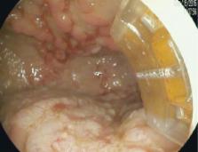
           Gastric antral vascular ectasia (GAVE) is a mucosal abnormality associated with multiple conditions, most notably cirrhosis and systemic sclerosis, that causes indolent gastrointestinal bleeding. It is primarily managed with endoscopic therapy. Traditionally, GAVE is endoscopically ablated using argon plasma coagulation (APC) but radiofrequency ablation (RFA) is emerging as an alternative modality. No prior comparison of the 2 modalities has been published.

          Patients and methods

           After receiving IRB approval, we reviewed our electronic health records to identify all patients who underwent endoscopic evaluation for GAVE between January, 2011 and October, 2016. We compared important variables between APC and RFA, as well as between cirrhosis and non-cirrhosis, using the Chi-square test and the Wilcoxon two-sample test as appropriate.

          Results

           During our study period, 81 patients were endoscopically evaluated for GAVE. 24 patients were treated with APC alone, 28 with RFA alone and 25 patients received both treatment modalities.

          Conclusions

           RFA and APC were both effective in treating bleeding from GAVE. Though we found subtle differences, patients underwent a similar number of treatment sessions and had similar procedure times and a similar time between sessions no matter the treatment modality used.

          Related collections

          Most cited references17

          • Record: found
          • Abstract: found
          • Article: not found

          Diagnosis and management of gastric antral vascular ectasia.

          Gastric antral vascular ectasia (GAVE) is an uncommon but often severe cause of upper gastrointestinal (GI) bleeding, responsible of about 4% of non-variceal upper GI haemorrhage. The diagnosis is mainly based on endoscopic pattern and, for uncertain cases, on histology. GAVE is characterized by a pathognomonic endoscopic pattern, mainly represented by red spots either organized in stripes radially departing from pylorus, defined as watermelon stomach, or arranged in a diffused-way, the so called honeycomb stomach. The histological pattern, although not pathognomonic, is characterized by four alterations: vascular ectasia of mucosal capillaries, focal thrombosis, spindle cell proliferation and fibrohyalinosis, which consist of homogeneous substance around the ectatic capillaries of the lamina propria. The main differential diagnosis is with Portal Hypertensive Gastropathy, that can frequently co-exists, since about 30% of patients with GAVE co-present a liver cirrhosis. Autoimmune disorders, mainly represented by Reynaud's phenomenon and sclerodactyly, are co-present in about 60% of patients with GAVE; other autoimmune and connective tissue disorders are occasionally reported such as Sjogren's syndrome, systemic lupus erythematosus, primary biliary cirrhosis and systemic sclerosis. In the remaining cases, GAVE syndrome has been described in patients with chronic renal failure, bone marrow transplantation and cardiac diseases. The pathogenesis of GAVE is still obscure and many hypotheses have been proposed such as mechanical stress, humoural and autoimmune factors and hemodynamic alterations. In the last two decades, many therapeutic options have been proposed including surgical, endoscopic and medical choices. Medical therapy has not clearly shown satisfactory results and surgery should only be considered for refractory severe cases, since this approach has significant mortality and morbidity risks, especially in the setting of portal hypertension and liver cirrhosis. Endoscopic therapy, particularly treatment with Argon Plasma Coagulation, has shown to be as effective and also safer than surgery, and should be considered the first-line treatment for patients with GAVE-related bleeding.
            Bookmark
            • Record: found
            • Abstract: found
            • Article: found

            Gastric Antral Vascular Ectasia (GAVE): An Update on Clinical Presentation, Pathophysiology and Treatment

            Gastric antral vascular ectasia (GAVE), though a rare disorder, causes up to 4% of non-variceal upper GI bleeding. This paper gives an overview of studies examining clinical presentation and pathophysiology, and reviews the current evidence for invasive and non-invasive treatments. GAVE is often associated with systemic illnesses, such as cirrhosis of the liver, autoimmune connective tissue disorders, bone marrow transplantation and chronic renal failure. The pathophysiological changes leading to GAVE have not been fully explained and remain controversial. Patient presentation varies from chronic iron-deficiency anaemia to heavy acute gastrointestinal bleeding. It is important to differentiate GAVE from portal hypertensive gastropathy as GAVE does not respond to measures reducing portal pressures. Endoscopic ablation (Nd:YAG-laser or argon plasma coagulation) is the first-line treatment of choice. As evidence for pharmacological therapy with oestrogen (and/or progesterone), tranexamic acid or thalidomide stems from case reports only, these should be used if endoscopic measures have failed to stop chronic blood loss. Surgical antrectomy should be reserved for unresponsive cases as it is associated with a high mortality. Ultimately, treatment of the underlying medical co-morbidities may lead to resolution of GAVE.
              Bookmark
              • Record: found
              • Abstract: found
              • Article: not found

              The clinical and endoscopic spectrum of the watermelon stomach.

              The watermelon stomach is an uncommon but treatable cause of chronic gastrointestinal bleeding. We report our experience with the clinical and endoscopic features of 45 consecutive patients treated by endoscopic Nd:YAG laser coagulation. The prototypic patient was a woman (71%) with an average age of 73 years (range of 53-89 years) who presented with occult (89%) transfusion-dependent (62%) gastrointestinal bleeding over a median period of 2 years (range of 1 month to > 20 years). Autoimmune connective tissue disorders were present in 28 patients (62%), especially Raynaud's phenomena (31%) and sclerodactyly (20%). Atrophic gastritis occurred in 19 of 19 (100%) patients, with hypergastrinemia in 25 (76%) of 33 patients tested. Antral endoscopic appearances included raised or flat stripes of ectatic vascular tissue (89%) or diffusely scattered lesions (11%). Proximal gastric involvement was present in 12 patients (27%), typically in the presence of a diaphragmatic hernia. Endoscopic laser therapy after a median of one treatment (range of 1-4) resulted in complete resolution of visible disease in four patients (13%) and resolution of > 90% in 24 patients (80%). Hemoglobin levels normalized in 87% of patients over a median follow-up period of 2 years (range of 1 month to 6 years) with no major complications. Blood transfusions were not necessary after laser therapy in 86% of 28 initially transfusion-dependent patients. The characteristic clinical, laboratory, and endoscopic features allow for a confident diagnosis that can lead to successful endoscopic treatment.
                Bookmark

                Author and article information

                Journal
                Endosc Int Open
                Endosc Int Open
                10.1055/s-00025476
                Endoscopy International Open
                © Georg Thieme Verlag KG (Stuttgart · New York )
                2364-3722
                2196-9736
                March 2018
                28 February 2018
                : 6
                : 3
                : E266-E270
                Affiliations
                [1 ]Division of Gastroenterology, Duke University Medical Center, Durham, North Carolina, United States
                [2 ]Division of Internal Medicine, Duke University Medical Center, Durham, North Carolina, United States
                [3 ]Division of Biostatistics, Duke University Medical Center, Durham, North Carolina, United States
                Author notes
                Corresponding author Paul St. Romain Duke University Medical Center Department of Medicine, Division of Gastroenterology 03142 Orange ZoneDurham, NC 27710+1-919-684-8857 Paul.st.romain@ 123456duke.edu
                Article
                10.1055/s-0043-123187
                5829995
                29497685
                189d465e-6cba-41f8-86ee-ff7abce106ae

                This is an open-access article distributed under the terms of the Creative Commons Attribution-NonCommercial-NoDerivatives License, which permits unrestricted reproduction and distribution, for non-commercial purposes only; and use and reproduction, but not distribution, of adapted material for non-commercial purposes only, provided the original work is properly cited.

                History
                : 25 August 2017
                : 08 November 2017
                Categories
                Original article

                Comments

                Comment on this article