21
views
0
recommends
+1 Recommend
0 collections
    0
    shares
      • Record: found
      • Abstract: found
      • Article: found
      Is Open Access

      Right ventricular mechanical pattern in health and disease: beyond longitudinal shortening

      review-article

      Read this article at

      Bookmark
          There is no author summary for this article yet. Authors can add summaries to their articles on ScienceOpen to make them more accessible to a non-specialist audience.

          Abstract

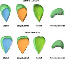
          Right ventricular (RV) function has proven to be a prognostic factor in heart failure with reduced and preserved ejection fraction and in pulmonary hypertension. RV function is also a cornerstone in the management of novel clinical issues, such as mechanical circulatory support devices or grown-up congenital heart disease patients. Despite the notable amount of circumferentially oriented myofibers in the subepicardial layer of the RV myocardium, the non-longitudinal motion directions are often neglected in the everyday assessment of RV function by echocardiography. However, the complex RV contraction pattern incorporates different motion components along three anatomically relevant axes: longitudinal shortening with traction of the tricuspid annulus towards the apex, radial motion of free wall often referred as the “bellows effect”, and anteroposterior shortening of the chamber by stretching the free wall over the septum. Advanced echocardiographic techniques, such as speckle-tracking and 3D echocardiography allow an in-depth characterization of RV mechanical pattern, providing better understanding of RV systolic and diastolic function. In our current review, we summarize the existing knowledge regarding RV mechanical adaptation to pressure- and/or volume-overloaded states and also other physiologic or pathologic conditions.

          Related collections

          Most cited references43

          • Record: found
          • Abstract: found
          • Article: found
          Is Open Access

          Repaired tetralogy of Fallot: the roles of cardiovascular magnetic resonance in evaluating pathophysiology and for pulmonary valve replacement decision support

          Tal Geva (2011)
          Surgical management of tetralogy of Fallot (TOF) results in anatomic and functional abnormalities in the majority of patients. Although right ventricular volume load due to severe pulmonary regurgitation can be tolerated for many years, there is now evidence that the compensatory mechanisms of the right ventricular myocardium ultimately fail and that if the volume load is not eliminated or reduced by pulmonary valve replacement the dysfunction might be irreversible. Cardiovascular magnetic resonance (CMR) has evolved during the last 2 decades as the reference standard imaging modality to assess the anatomic and functional sequelae in patients with repaired TOF. This article reviews the pathophysiology of chronic right ventricular volume load after TOF repair and the risks and benefits of pulmonary valve replacement. The CMR techniques used to comprehensively evaluate the patient with repaired TOF are reviewed and the role of CMR in supporting clinical decisions regarding pulmonary valve replacement is discussed.
            Bookmark
            • Record: found
            • Abstract: found
            • Article: not found

            Right heart dysfunction and failure in heart failure with preserved ejection fraction: mechanisms and management. Position statement on behalf of the Heart Failure Association of the European Society of Cardiology

            There is an unmet need for effective treatment strategies to reduce morbidity and mortality in patients with heart failure with preserved ejection fraction (HFpEF). Until recently, attention in patients with HFpEF was almost exclusively focused on the left side. However, it is now increasingly recognized that right heart dysfunction is common and contributes importantly to poor prognosis in HFpEF. More insights into the development of right heart dysfunction in HFpEF may aid to our knowledge about this complex disease and may eventually lead to better treatments to improve outcomes in these patients. In this position paper from the Heart Failure Association of the European Society of Cardiology, the Committee on Heart Failure with Preserved Ejection Fraction reviews the prevalence, diagnosis, and pathophysiology of right heart dysfunction and failure in patients with HFpEF. Finally, potential treatment strategies, important knowledge gaps and future directions regarding the right side in HFpEF are discussed.
              Bookmark
              • Record: found
              • Abstract: found
              • Article: not found

              Exercise-induced pulmonary hypertension: physiological basis and methodological concerns.

              Exercise stresses the pulmonary circulation through increases in cardiac output (.Q) and left atrial pressure. Invasive as well as noninvasive studies in healthy volunteers show that the slope of mean pulmonary artery pressure (mPAP)-flow relationships ranges from 0.5 to 3 mm Hg.min.L(-1). The upper limit of normal mPAP at exercise thus approximates 30 mm Hg at a .Q of less than 10 L.min(-1) or a total pulmonary vascular resistance at exercise of less than 3 Wood units. Left atrial pressure increases at exercise with an average upstream transmission to PAP in a close to one-for-one mm Hg fashion. Multipoint PAP-flow relationships are usually described by a linear approximation, but present with a slight curvilinearity, which is explained by resistive vessel distensibility. When mPAP is expressed as a function of oxygen uptake or workload, plateau patterns may be observed in patients with systolic heart failure who cannot further increase .Q at the highest levels of exercise. Exercise has to be dynamic to avoid the increase in systemic vascular resistance and abrupt changes in intrathoracic pressure that occur with resistive exercise and can lead to unpredictable effects on the pulmonary circulation. Postexercise measurements are unreliable because of the rapid return of pulmonary vascular pressures and flows to the baseline resting state. Recent studies suggest that exercise-induced increase in PAP to a mean higher than 30 mm Hg may be associated with dyspnea-fatigue symptomatology.
                Bookmark

                Author and article information

                Contributors
                +36206663427 , attila.kovacs@med.semmelweis-univ.hu
                Journal
                Heart Fail Rev
                Heart Fail Rev
                Heart Failure Reviews
                Springer US (New York )
                1382-4147
                1573-7322
                9 March 2019
                9 March 2019
                2019
                : 24
                : 4
                : 511-520
                Affiliations
                ISNI 0000 0001 0942 9821, GRID grid.11804.3c, Semmelweis University Heart and Vascular Center, ; Városmajor St. 68, Budapest, H-1122 Hungary
                Author information
                http://orcid.org/0000-0003-2320-6434
                Article
                9778
                10.1007/s10741-019-09778-1
                6559995
                30852772
                18309286-46ce-4b5f-8a1d-dbd52e1bb871
                © The Author(s) 2019

                Open Access This article is distributed under the terms of the Creative Commons Attribution 4.0 International License (http://creativecommons.org/licenses/by/4.0/), which permits unrestricted use, distribution, and reproduction in any medium, provided you give appropriate credit to the original author(s) and the source, provide a link to the Creative Commons license, and indicate if changes were made.

                History
                Funding
                Funded by: Semmelweis University (SE)
                Categories
                Article
                Custom metadata
                © Springer Science+Business Media, LLC, part of Springer Nature 2019

                Cardiovascular Medicine
                right ventricle,3d echocardiography,speckle tracking,heart failure,pulmonary hypertension

                Comments

                Comment on this article