7
views
0
recommends
+1 Recommend
0 collections
    0
    shares
      • Record: found
      • Abstract: found
      • Article: found
      Is Open Access

      CTNND1 variants cause familial exudative vitreoretinopathy through the Wnt/cadherin axis

      research-article

      Read this article at

      Bookmark
          There is no author summary for this article yet. Authors can add summaries to their articles on ScienceOpen to make them more accessible to a non-specialist audience.

          Abstract

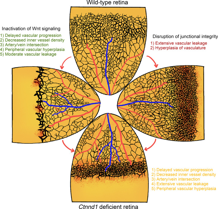
          Familial exudative vitreoretinopathy (FEVR) is a hereditary disorder that can cause vision loss. CTNND1 encodes a cellular adhesion protein p120-catenin (p120), which is essential for vascularization with unclear function in postnatal physiological angiogenesis. Here, we applied whole-exome sequencing to 140 probands of FEVR families and identified 3 candidate variants in the human CTNND1 gene. We performed inducible deletion of Ctnnd1 in the postnatal mouse endothelial cells (ECs) and observed typical phenotypes of FEVR with reactive gliosis. Using unbiased proteomics analysis combined with experimental approaches, we conclude that p120 is critical for the integrity of adherens junctions (AJs) and that p120 activates Wnt signaling activity by protecting β-catenin from glycogen synthase kinase 3 beta–ubiqutin–guided (Gsk3β-ubiquitin–guided) degradation. Treatment of CTNND1-depleted human retinal microvascular ECs with Gsk3β inhibitors LiCl or CHIR-99021 enhanced cell proliferation. Moreover, LiCl treatment increased vessel density in Ctnnd1-deficient mouse retinas. Variants in CTNND1 caused FEVR by compromising the expression of AJs and Wnt signaling activity. Genetic interactions between p120 and β-catenin or α-catenin revealed by double-heterozygous deletion in mice showed that p120 regulates vascular development through the Wnt/cadherin axis. In conclusion, variants in CTNND1 can cause FEVR through the Wnt/cadherin axis.

          Abstract
          Related collections

          Most cited references73

          • Record: found
          • Abstract: found
          • Article: not found

          Wnt/β-Catenin Signaling, Disease, and Emerging Therapeutic Modalities.

          The WNT signal transduction cascade is a main regulator of development throughout the animal kingdom. Wnts are also key drivers of most types of tissue stem cells in adult mammals. Unsurprisingly, mutated Wnt pathway components are causative to multiple growth-related pathologies and to cancer. Here, we describe the core Wnt/β-catenin signaling pathway, how it controls stem cells, and contributes to disease. Finally, we discuss strategies for Wnt-based therapies.
            Bookmark
            • Record: found
            • Abstract: found
            • Article: found
            Is Open Access

            A Computational Tool for Quantitative Analysis of Vascular Networks

            Angiogenesis is the generation of mature vascular networks from pre-existing vessels. Angiogenesis is crucial during the organism' development, for wound healing and for the female reproductive cycle. Several murine experimental systems are well suited for studying developmental and pathological angiogenesis. They include the embryonic hindbrain, the post-natal retina and allantois explants. In these systems vascular networks are visualised by appropriate staining procedures followed by microscopical analysis. Nevertheless, quantitative assessment of angiogenesis is hampered by the lack of readily available, standardized metrics and software analysis tools. Non-automated protocols are being used widely and they are, in general, time - and labour intensive, prone to human error and do not permit computation of complex spatial metrics. We have developed a light-weight, user friendly software, AngioTool, which allows for quick, hands-off and reproducible quantification of vascular networks in microscopic images. AngioTool computes several morphological and spatial parameters including the area covered by a vascular network, the number of vessels, vessel length, vascular density and lacunarity. In addition, AngioTool calculates the so-called “branching index” (branch points / unit area), providing a measurement of the sprouting activity of a specimen of interest. We have validated AngioTool using images of embryonic murine hindbrains, post-natal retinas and allantois explants. AngioTool is open source and can be downloaded free of charge.
              Bookmark
              • Record: found
              • Abstract: found
              • Article: not found

              The many faces and functions of β-catenin.

              β-Catenin (Armadillo in Drosophila) is a multitasking and evolutionary conserved molecule that in metazoans exerts a crucial role in a multitude of developmental and homeostatic processes. More specifically, β-catenin is an integral structural component of cadherin-based adherens junctions, and the key nuclear effector of canonical Wnt signalling in the nucleus. Imbalance in the structural and signalling properties of β-catenin often results in disease and deregulated growth connected to cancer and metastasis. Intense research into the life of β-catenin has revealed a complex picture. Here, we try to capture the state of the art: we try to summarize and make some sense of the processes that regulate β-catenin, as well as the plethora of β-catenin binding partners. One focus will be the interaction of β-catenin with different transcription factors and the potential implications of these interactions for direct cross-talk between β-catenin and non-Wnt signalling pathways.
                Bookmark

                Author and article information

                Contributors
                Journal
                JCI Insight
                JCI Insight
                JCI Insight
                JCI Insight
                American Society for Clinical Investigation
                2379-3708
                22 July 2022
                22 July 2022
                22 July 2022
                : 7
                : 14
                : e158428
                Affiliations
                [1 ]Sichuan Provincial Key Laboratory for Human Disease Gene Study, Center for Medical Genetics, Sichuan Provincial People’s Hospital, University of Electronic Science and Technology of China, Chengdu, China.
                [2 ]Research Unit for Blindness Prevention of the Chinese Academy of Medical Sciences (2019RU026), Sichuan Academy of Medical Sciences and Sichuan Provincial People’s Hospital, Chengdu, China.
                [3 ]State Key Laboratory of Ophthalmology, Zhongshan Ophthalmic Center, Sun Yat-sen University, Guangzhou, China.
                [4 ]Department of Ophthalmology, Xinhua Hospital Affiliated to Shanghai Jiaotong University School of Medicine, Shanghai, China.
                [5 ]Key Laboratory of Tibetan Medicine Research and Qinghai Provincial Key Laboratory of Tibetan Medicine Research, Northwest Institute of Plateau Biology, Chinese Academy of Sciences, Xining, China.
                Author notes
                Address correspondence to: Zhenglin Yang or Xianjun Zhu, No. 32 W. Sec. 2, 1st Ring Rd., 610072 Chengdu, Sichuan, China. Phone: 86.02887393375; Email: yangzhenglin@ 123456cashq.ac.cn (ZY); Email: xjzhu@ 123456uestc.edu.cn (XZ). Or to: Xiaoyan Ding, No. 135, Xingang Xi Road, 510275 Guangzhou, China. Phone: 86.02887393375; Email: dingxiaoyan@ 123456gzzoc.com .

                Authorship note: MY, SL, LH, and RZ contributed equally to this work.

                Author information
                http://orcid.org/0000-0002-6001-6827
                http://orcid.org/0000-0002-7691-3776
                http://orcid.org/0000-0002-9434-7499
                http://orcid.org/0000-0002-5265-3244
                http://orcid.org/0000-0002-5288-3816
                http://orcid.org/0000-0002-2531-7552
                Article
                158428
                10.1172/jci.insight.158428
                9431724
                35700046
                1810c675-db24-438f-b034-53170b09e625
                © 2022 Yang et al.

                This work is licensed under the Creative Commons Attribution 4.0 International License. To view a copy of this license, visit http://creativecommons.org/licenses/by/4.0/.

                History
                : 12 January 2022
                : 10 June 2022
                Funding
                Funded by: National Natural Science Foundation of China, https://doi.org/10.13039/501100001809;
                Award ID: 82101153,82000913,81970841,81790643,82121003
                Funded by: Department of Science and Technology of Sichuan Province, https://doi.org/10.13039/501100004829;
                Award ID: 2021YFS0386,2021YFS0369,2021JDGD0036
                Funded by: he CAMS Innovation Fund for Medical Sciences
                Award ID: 2019-12M-5-032
                Funded by: the fund for Sichuan Provincial People’s Hospital
                Award ID: 2021QN01
                Funded by: Department of Chengdu Science and Technology
                Award ID: 2021-YF05-01316-SN
                Categories
                Research Article

                genetics,ophthalmology,endothelial cells,genetic diseases

                Comments

                Comment on this article