4
views
0
recommends
+1 Recommend
0 collections
    0
    shares
      • Record: found
      • Abstract: found
      • Article: found
      Is Open Access

      Simplifying the Surgical Classification and Approach to the Posterolateral Skull Base and Jugular Foramen Using Anatomical Triangles

      research-article

      Read this article at

      Bookmark
          There is no author summary for this article yet. Authors can add summaries to their articles on ScienceOpen to make them more accessible to a non-specialist audience.

          Abstract

          Introduction

          Lesions of the jugular foramen (JF) and postero-lateral skull base are difficult to expose and exhibit complex neurovascular relationships. Given their rarity and the increasing use of radiosurgery, neurosurgeons are becoming less experienced with their surgical management. Anatomical factors are crucial in designing the approach to achieve a maximal safe resection.

          Methods and methods

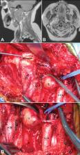
          Six cadaveric heads (12 sides) were dissected via combined post-auricular infralabyrinthine and distal transcervical approach with additional anterior transstyloid and posterior far lateral exposures. Contiguous surgical triangles were measured, and contents were analyzed. Thirty-one patients (32 lesions) were treated surgically between 2000 and 2016 through different variations of the retro-auricular distal cervical transtemporal approaches.

          Results

          We anatomically reviewed the carotid, stylodigastric, jugular, condylar, suboccipital, deep condylar, mastoid, suprajugular, suprahypoglossal (infrajugular), and infrahypoglossal triangles. Tumors included glomus jugulare, lower cranial nerve schwannomas or neurofibromas, meningiomas, chondrosarcoma, adenocystic carcinoma, plasmacytoma of the occipitocervical joint, and a sarcoid lesion. We classified tumors into extracranial, intradural, intraosseous, and dumbbell-shaped, and analyzed the approach selection for each.

          Conclusion

          Jugular foramen and posterolateral skull base lesions can be safely resected through a retro-auricular distal cervical lateral skull base approach, which is customizable to anatomical location and tumor extension by tailoring the involved osteo-muscular triangles.

          Related collections

          Most cited references20

          • Record: found
          • Abstract: found
          • Article: not found

          Microsurgical anatomy of the transcondylar, supracondylar, and paracondylar extensions of the far-lateral approach.

          Despite a large number of reports of the use of the far-lateral approach, some of the basic detail that is important in safely completing this exposure has not been defined or remains poorly understood. The basic far-lateral exposure provides access for the following approaches: 1) the transcondylar approach directed through the occipital condyle or the adjoining portions of the occipital and atlantal condyles; 2) the supracondylar approach directed through the area above the occipital condyle; and 3) the paracondylar exposure directed through the area lateral to the occipital condyle. The transcondylar approach provides access to the lower clivus and premedullary area. The supracondylar approach provides access to the region of, and medial to, the hypoglossal canal and jugular tubercle. The paracondylar approach, which includes drilling of the jugular process of the occipital bone in the area lateral to the occipital condyle, provides access to the posterior portion of the jugular foramen and to the mastoid on the lateral side of the jugular foramen. In this study, the anatomy important to completing the far-lateral approach and these modifications was examined in 12 cadaveric specimens. In the standard posterior and posterolateral approaches, an understanding of the individual suboccipital muscles is not essential. However, these muscles provide important landmarks for the far-lateral approach and its modifications. Other important considerations include the relationship of the occipital condyle to the foramen magnum, hypoglossal canal, jugular tubercle, the jugular process of the occipital bone, the mastoid, and the facial canal. These and other relationships important to completing these exposures were examined in this study.
            Bookmark
            • Record: found
            • Abstract: found
            • Article: not found

            Lateral suboccipital approach for vertebral and vertebrobasilar artery lesions.

            R Heros (1986)
            A modification of the unilateral suboccipital approach is elaborated and illustrated. This modification is useful for aneurysms of the vertebral artery, the vertebrobasilar junction, and the proximal basilar trunk, and for arteriovenous malformations of the inferolateral cerebellum. It entails extreme lateral removal of the rim of the foramen magnum toward the condylar fossa and posterolateral removal of the arch of the atlas toward the exposed vertebral artery. This extra bone removal allows an approach to the front of the brain stem from inferolaterally, after gentle upward and medial retraction of the tonsil, with minimal or no retraction of the medulla.
              Bookmark
              • Record: found
              • Abstract: found
              • Article: not found

              Subtemporal-preauricular infratemporal fossa approach to large lateral and posterior cranial base neoplasms.

              A subtemporal-preauricular infratemporal fossa approach to remove 22 large neoplasms involving the lateral and posterior cranial base is detailed. The areas from which a neoplasm could be removed by this approach included the sphenoid and clival bone; the medial half of the petrous temporal bone; the infratemporal fossa; the nasopharynx; the retro- and parapharyngeal area; the ethmoid, sphenoid, and maxillary sinuses; and the intradural clivus-foramen magnum area. The pathology of the neoplasms included benign tumors such as meningioma, malignant cartilaginous neoplasms such as chordoma, and other malignant lesions such as nasopharyngeal carcinoma. This approach offers many advantages over other anterior and lateral approaches to the lateral and posterior cranial base: these include minimal brain retraction; direct access to the ipsilateral petrous and upper cervical internal carotid artery; reconstruction of extensive cranial base defects, often with the use of a vascularized rectus abdominus flap; preservation of the hearing conduction mechanism when it is not involved by tumor; and the maintenance of excellent facial nerve function postoperatively. The use of an anterior extradural approach (transethmoidal) and of an intradural approach (frontotemporal or retromastoid), either concurrently or separately, is necessary in some patients to effect total tumor removal. The most serious complication in this series was the death of a patient due to postoperative infection and bilateral carotid artery rupture, which may have been avoided by the use of a rectus abdominis muscle flap for reconstruction. Among the 21 surviving patients, 18 had a good outcome, two had a fair outcome, and one with preexisting neurological deficits had a poor outcome. One of the surviving patients with a chordoma died of pulmonary metastases 1 year later, without evidence of local recurrence. The length of postoperative follow-up evaluation in these patients is insufficient to make any judgment about the effectiveness of this surgical approach in achieving a cure or long-term control of the tumors described.
                Bookmark

                Author and article information

                Journal
                Cureus
                Cureus
                2168-8184
                Cureus
                Cureus (Palo Alto (CA) )
                2168-8184
                16 November 2021
                November 2021
                : 13
                : 11
                : e19638
                Affiliations
                [1 ] Neurological Surgery, The University of Tennessee Health Science Center, Memphis, USA
                [2 ] Laboratory, Medical Education Research Institute, Memphis, USA
                [3 ] Neurosurgery, The University of Tennessee Health Science Center, Memphis, USA
                [4 ] Neurological Surgery, Semmes-Murphey Clinic, Memphis, USA
                Author notes
                Article
                10.7759/cureus.19638
                8676706
                1807e6d7-4961-498b-94e4-866d95a568c9
                Copyright © 2021, Basma et al.

                This is an open access article distributed under the terms of the Creative Commons Attribution License, which permits unrestricted use, distribution, and reproduction in any medium, provided the original author and source are credited.

                History
                : 16 November 2021
                Categories
                Neurosurgery
                Anatomy

                meningioma,glomus jugulare,hypoglossal schwannoma,distal cervical approach,hypoglossal canal,styloid process,infralabyrinthine approach,far lateral,jugular foramen

                Comments

                Comment on this article