22
views
0
recommends
+1 Recommend
0 collections
    0
    shares
      • Record: found
      • Abstract: found
      • Article: found
      Is Open Access

      Cdc42 regulates junctional actin but not cell polarization in the Caenorhabditis elegans epidermis

      research-article

      Read this article at

      Bookmark
          There is no author summary for this article yet. Authors can add summaries to their articles on ScienceOpen to make them more accessible to a non-specialist audience.

          Abstract

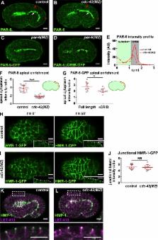
          Adherens junction remodeling allows changes in cell shape and position. Zilberman et al. show through live imaging that CDC-42 is dispensable for epithelial cell polarization, but its RhoGAP-regulated activity is needed to control junctional actin organization during embryo elongation.

          Abstract

          During morphogenesis, adherens junctions (AJs) remodel to allow changes in cell shape and position while preserving adhesion. Here, we examine the function of Rho guanosine triphosphatase CDC-42 in AJ formation and regulation during Caenorhabditis elegans embryo elongation, a process driven by asymmetric epidermal cell shape changes. cdc-42 mutant embryos arrest during elongation with epidermal ruptures. Unexpectedly, we find using time-lapse fluorescence imaging that cdc-42 is not required for epidermal cell polarization or junction assembly, but rather is needed for proper junctional actin regulation during elongation. We show that the RhoGAP PAC-1/ARHGAP21 inhibits CDC-42 activity at AJs, and loss of PAC-1 or the interacting linker protein PICC-1/CCDC85A-C blocks elongation in embryos with compromised AJ function. pac-1 embryos exhibit dynamic accumulations of junctional F-actin and an increase in AJ protein levels. Our findings identify a previously unrecognized molecular mechanism for inhibiting junctional CDC-42 to control actin organization and AJ protein levels during epithelial morphogenesis.

          Related collections

          Most cited references85

          • Record: found
          • Abstract: found
          • Article: not found

          Efficient gene transfer in C.elegans: extrachromosomal maintenance and integration of transforming sequences.

          We describe a dominant behavioral marker, rol-6(su-1006), and an efficient microinjection procedure which facilitate the recovery of Caenorhabditis elegans transformants. We use these tools to study the mechanism of C.elegans DNA transformation. By injecting mixtures of genetically marked DNA molecules, we show that large extrachromosomal arrays assemble directly from the injected molecules and that homologous recombination drives array assembly. Appropriately placed double-strand breaks stimulated homologous recombination during array formation. Our data indicate that the size of the assembled transgenic structures determines whether or not they will be maintained extrachromosomally or lost. We show that low copy number extrachromosomal transformation can be achieved by adjusting the relative concentration of DNA molecules in the injection mixture. Integration of the injected DNA, though relatively rare, was reproducibly achieved when single-stranded oligonucleotide was co-injected with the double-stranded DNA.
            Bookmark
            • Record: found
            • Abstract: not found
            • Article: not found

            Specific interference by ingested dsRNA.

              Bookmark
              • Record: found
              • Abstract: found
              • Article: not found

              PTEN-mediated apical segregation of phosphoinositides controls epithelial morphogenesis through Cdc42.

              Formation of the apical surface and lumen is a fundamental, yet poorly understood, step in epithelial organ development. We show that PTEN localizes to the apical plasma membrane during epithelial morphogenesis to mediate the enrichment of PtdIns(4,5)P2 at this domain during cyst development in three-dimensional culture. Ectopic PtdIns(4,5)P2 at the basolateral surface causes apical proteins to relocalize to the basolateral surface. Annexin 2 (Anx2) binds PtdIns(4,5)P2 and is recruited to the apical surface. Anx2 binds Cdc42, recruiting it to the apical surface. Cdc42 recruits aPKC to the apical surface. Loss of function of PTEN, Anx2, Cdc42, or aPKC prevents normal development of the apical surface and lumen. We conclude that the mechanism of PTEN, PtdIns(4,5)P2, Anx2, Cdc42, and aPKC controls apical plasma membrane and lumen formation.
                Bookmark

                Author and article information

                Journal
                J Cell Biol
                J. Cell Biol
                jcb
                jcb
                The Journal of Cell Biology
                The Rockefeller University Press
                0021-9525
                1540-8140
                06 November 2017
                : 216
                : 11
                : 3729-3744
                Affiliations
                [1 ]Helen L. and Martin S. Kimmel Center for Biology and Medicine at the Skirball Institute of Biomolecular Medicine, New York University School of Medicine, New York, NY
                [2 ]Department of Cell Biology, New York University School of Medicine, New York, NY
                Author notes
                Correspondence to Jeremy Nance: jeremy.nance@ 123456med.nyu.edu
                Author information
                http://orcid.org/0000-0003-4212-7731
                Article
                201611061
                10.1083/jcb.201611061
                5674880
                28903999
                169ba87e-884d-467e-ab72-1ed767bad47b
                © 2017 Zilberman et al.

                This article is distributed under the terms of an Attribution–Noncommercial–Share Alike–No Mirror Sites license for the first six months after the publication date (see http://www.rupress.org/terms/). After six months it is available under a Creative Commons License (Attribution–Noncommercial–Share Alike 4.0 International license, as described at https://creativecommons.org/licenses/by-nc-sa/4.0/).

                History
                : 10 November 2016
                : 18 July 2017
                : 15 August 2017
                Funding
                Funded by: National Institutes of Health, DOI http://dx.doi.org/10.13039/100000002;
                Award ID: P40 OD010440
                Funded by: National Institutes of Health, DOI http://dx.doi.org/10.13039/100000002;
                Award ID: R01GM098492
                Award ID: R01GM078341
                Award ID: R35GM118081
                Funded by: American Hospital Association, DOI http://dx.doi.org/10.13039/100008438;
                Award ID: 14POST18740059
                Categories
                Research Articles
                Article
                20
                38

                Cell biology
                Cell biology

                Comments

                Comment on this article