6
views
0
recommends
+1 Recommend
0 collections
    0
    shares
      • Record: found
      • Abstract: found
      • Article: found
      Is Open Access

      Rosette-forming glioneuronal tumours: two case reports and a review of the literature

      case-report
      , MD 1 , , MD 1 , , MD 1 , , MD 1 , , MD, PhD 1 ,
      BJR | Case Reports
      The British Institute of Radiology.

      Read this article at

      Bookmark
          There is no author summary for this article yet. Authors can add summaries to their articles on ScienceOpen to make them more accessible to a non-specialist audience.

          Abstract

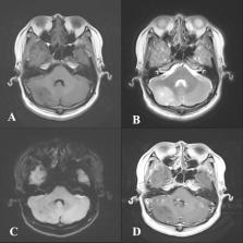
          Rosette-forming glioneuronal tumour (RGNT) is a rare central nervous system (CNS) neoplasm that typically arises in the fourth ventricle. It is even more uncommon to arise outside the midline. In this paper, we report two cases of RGNT: one located in the fourth ventricle (a typical site), and the other in the right cerebellar hemisphere (a rare site). Both cases were misdiagnosed on imaging, and the results were inconsistent with the pathological diagnosis. The aim of the article is to deepen medical practitioners’ understanding of RGNT by learning from these two cases, summarising cases located in the cerebellar hemispheres and systematically reviewing RGNT.

          Related collections

          Most cited references16

          • Record: found
          • Abstract: found
          • Article: found

          The 2016 World Health Organization Classification of Tumors of the Central Nervous System: a summary.

          The 2016 World Health Organization Classification of Tumors of the Central Nervous System is both a conceptual and practical advance over its 2007 predecessor. For the first time, the WHO classification of CNS tumors uses molecular parameters in addition to histology to define many tumor entities, thus formulating a concept for how CNS tumor diagnoses should be structured in the molecular era. As such, the 2016 CNS WHO presents major restructuring of the diffuse gliomas, medulloblastomas and other embryonal tumors, and incorporates new entities that are defined by both histology and molecular features, including glioblastoma, IDH-wildtype and glioblastoma, IDH-mutant; diffuse midline glioma, H3 K27M-mutant; RELA fusion-positive ependymoma; medulloblastoma, WNT-activated and medulloblastoma, SHH-activated; and embryonal tumour with multilayered rosettes, C19MC-altered. The 2016 edition has added newly recognized neoplasms, and has deleted some entities, variants and patterns that no longer have diagnostic and/or biological relevance. Other notable changes include the addition of brain invasion as a criterion for atypical meningioma and the introduction of a soft tissue-type grading system for the now combined entity of solitary fibrous tumor / hemangiopericytoma-a departure from the manner by which other CNS tumors are graded. Overall, it is hoped that the 2016 CNS WHO will facilitate clinical, experimental and epidemiological studies that will lead to improvements in the lives of patients with brain tumors.
            Bookmark
            • Record: found
            • Abstract: found
            • Article: not found

            The 2007 WHO Classification of Tumours of the Central Nervous System

            The fourth edition of the World Health Organization (WHO) classification of tumours of the central nervous system, published in 2007, lists several new entities, including angiocentric glioma, papillary glioneuronal tumour, rosette-forming glioneuronal tumour of the fourth ventricle, papillary tumour of the pineal region, pituicytoma and spindle cell oncocytoma of the adenohypophysis. Histological variants were added if there was evidence of a different age distribution, location, genetic profile or clinical behaviour; these included pilomyxoid astrocytoma, anaplastic medulloblastoma and medulloblastoma with extensive nodularity. The WHO grading scheme and the sections on genetic profiles were updated and the rhabdoid tumour predisposition syndrome was added to the list of familial tumour syndromes typically involving the nervous system. As in the previous, 2000 edition of the WHO ‘Blue Book’, the classification is accompanied by a concise commentary on clinico-pathological characteristics of each tumour type. The 2007 WHO classification is based on the consensus of an international Working Group of 25 pathologists and geneticists, as well as contributions from more than 70 international experts overall, and is presented as the standard for the definition of brain tumours to the clinical oncology and cancer research communities world-wide.
              Bookmark
              • Record: found
              • Abstract: not found
              • Article: not found

              Rosette-forming glioneuronal tumors share a distinct DNA methylation profile and mutations in FGFR1, with recurrent co-mutation of PIK3CA and NF1

                Bookmark

                Author and article information

                Contributors
                Journal
                BJR Case Rep
                British Institute of Radiology
                bjrcr
                BJR | Case Reports
                The British Institute of Radiology.
                2055-7159
                01 January 2022
                26 November 2021
                : 8
                : 1
                : 20210125
                Affiliations
                [1 ]org-divisionDepartment of Radiology, The First Affiliated Hospital of Shenzhen University, Health Science Center, Shenzhen Second People’s Hospital , Shenzhen, China
                Author notes
                Address correspondence to: Fan Lin. E-mail: foxetfoxet@ 123456gmail.com
                Author information
                https://orcid.org/0000-0001-5731-7429
                Article
                BJRCR-D-21-00125
                10.1259/bjrcr.20210125
                8803231
                35136642
                16695238-d949-48da-bdd3-328d0ab9ef67
                © 2022 The Authors. Published by the British Institute of Radiology

                This is an open access article distributed under the terms of the Creative Commons Attribution 4.0 International License, which permits unrestricted use, distribution and reproduction in any medium, provided the original author and source are credited.

                History
                : 28 June 2021
                : 26 October 2021
                : 03 November 2021
                Page count
                Figures: 4, Tables: 1, Equations: 0, References: 16, Pages: 5, Words: 3174
                Categories
                Case Report
                bjrcr, BJRCR
                neuro, Neuroradiology

                Comments

                Comment on this article