4
views
0
recommends
+1 Recommend
0 collections
    0
    shares
      • Record: found
      • Abstract: found
      • Article: found
      Is Open Access

      Mex3a interacts with LAMA2 to promote lung adenocarcinoma metastasis via PI3K/AKT pathway

      research-article

      Read this article at

      Bookmark
          There is no author summary for this article yet. Authors can add summaries to their articles on ScienceOpen to make them more accessible to a non-specialist audience.

          Abstract

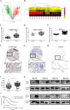
          Lung adenocarcinoma (LUAD) is the main subtype of lung cancer. In this study, we found that RBP Mex3a was significantly upregulated in LUAD tissues and elevated Mex3a expression was associated with poor LUAD prognosis and metastasis. Furthermore, we demonstrated that Mex3a knockdown significantly inhibited LUAD cell migration and invasion in vitro and metastasis in nude mice. Transcriptome sequencing indicated that Mex3a affected gene expression linked to ECM-receptor interactions, including laminin subunit alpha 2(LAMA2). RNA immunoprecipitation (RIP) assay revealed Mex3a directly bound to LAMA2 mRNA and Mex3a increased the instability of LAMA2 mRNA in LUAD cells. Furthermore, we discovered that LAMA2 was surprisingly downregulated in LUAD and inhibited LUAD metastasis. LAMA2 knockdown partially reverse the decrease of cell migration and invasion caused by Mex3a knockdown. In addition, we found that both Mex3a and LAMA2 could influence PI3K-AKT pathway, which are downstream effectors of the ECM-receptor pathway. Moreover, the reduced activation of PI3K-AKT pathway in caused by Mex3a depletion was rescued by LAMA2 knockdown. In conclusion, we demonstrated that Mex3a downregulates LAMA2 expression to exert a prometastatic role in LUAD. Our study revealed the prognostic and prometastatic effects of Mex3a in LUAD, suggesting that Mex3a can serve as a prognostic biomarker and a target for metastatic therapy.

          Related collections

          Most cited references21

          • Record: found
          • Abstract: found
          • Article: found
          Is Open Access

          Cell Adhesion and Matrix Stiffness: Coordinating Cancer Cell Invasion and Metastasis

          Metastasis is a multistep process in which tumor extracellular matrix (ECM) and cancer cell cytoskeleton interactions are pivotal. ECM is connected, through integrins, to the cell’s adhesome at cell–ECM adhesion sites and through them to the actin cytoskeleton and various downstream signaling pathways that enable the cell to respond to external stimuli in a coordinated manner. Cues from cell-adhesion proteins are fundamental for defining the invasive potential of cancer cells, and many of these proteins have been proposed as potent targets for inhibiting cancer cell invasion and thus, metastasis. In addition, ECM accumulation is quite frequent within the tumor microenvironment leading in many cases to an intense fibrotic response, known as desmoplasia, and tumor stiffening. Stiffening is not only required for the tumor to be able to displace the host tissue and grow in size but also contributes to cell–ECM interactions and can promote cancer cell invasion to surrounding tissues. Here, we review the role of cell adhesion and matrix stiffness in cancer cell invasion and metastasis.
            Bookmark
            • Record: found
            • Abstract: found
            • Article: not found

            Functional diversity of laminins.

            Laminins are a large family of conserved, multidomain trimeric basement membrane proteins that contribute to the structure of extracellular matrix and influence the behavior of associated cells, such as adhesion, differentiation, migration, phenotype stability, and resistance to anoikis. In lower organisms such as Hydra there is only one isoform of laminin, but higher organisms have at least 16 trimeric isoforms with varying degrees of cell/tissue specificity. In vitro protein and cell culture studies, gene manipulation in animals, and laminin gene mutations in human diseases have provided insight into the specific functions of some laminins, but the biological roles of many isoforms are still largely unexplored, mainly owing to difficulties in isolating them in pure form from tissues or cells. In this review, we elucidate the evolution of laminins, describe their molecular complexity, and explore the current knowledge of their diversity and functional aspects, including laminin-mediated signaling via membrane receptors, in vitro cell biology, and involvement in various tissues gained from animal model and human disease studies. The potential use of laminins in cell biology research and biotechnology is discussed.
              Bookmark
              • Record: found
              • Abstract: found
              • Article: not found

              Delineation of two clinically and molecularly distinct subgroups of posterior fossa ependymoma.

              Despite the histological similarity of ependymomas from throughout the neuroaxis, the disease likely comprises multiple independent entities, each with a distinct molecular pathogenesis. Transcriptional profiling of two large independent cohorts of ependymoma reveals the existence of two demographically, transcriptionally, genetically, and clinically distinct groups of posterior fossa (PF) ependymomas. Group A patients are younger, have laterally located tumors with a balanced genome, and are much more likely to exhibit recurrence, metastasis at recurrence, and death compared with Group B patients. Identification and optimization of immunohistochemical (IHC) markers for PF ependymoma subgroups allowed validation of our findings on a third independent cohort, using a human ependymoma tissue microarray, and provides a tool for prospective prognostication and stratification of PF ependymoma patients. Copyright © 2011 Elsevier Inc. All rights reserved.
                Bookmark

                Author and article information

                Contributors
                zuimeidexiaoyu@126.com
                tianhuiql@126.com
                Journal
                Cell Death Dis
                Cell Death Dis
                Cell Death & Disease
                Nature Publishing Group UK (London )
                2041-4889
                13 August 2020
                13 August 2020
                August 2020
                : 11
                : 8
                : 614
                Affiliations
                [1 ]GRID grid.27255.37, ISNI 0000 0004 1761 1174, Department of Thoracic Surgery, Qilu Hospital, Cheeloo College of Medicine, , Shandong University, ; 250012 Jinan, Shandong China
                [2 ]GRID grid.27255.37, ISNI 0000 0004 1761 1174, School of Basic Medical Sciences of Shandong University, ; 250012 Jinan, China
                [3 ]GRID grid.416966.a, ISNI 0000 0004 1758 1470, Weifang People’s Hospital, ; 261000 Weifang, China
                [4 ]GRID grid.27255.37, ISNI 0000 0004 1761 1174, Department of General Surgery, Cheeloo College of Medicine, , Shandong University, ; 250012 Jinan, Shandong China
                [5 ]GRID grid.27255.37, ISNI 0000 0004 1761 1174, School of Medicine, , Shandong University, ; 250012 Jinan, China
                Author information
                http://orcid.org/0000-0003-0568-0036
                http://orcid.org/0000-0001-5913-4781
                Article
                2858
                10.1038/s41419-020-02858-3
                7427100
                32792503
                1657d884-2422-497c-a099-7ef2857acbd0
                © The Author(s) 2020

                Open Access This article is licensed under a Creative Commons Attribution 4.0 International License, which permits use, sharing, adaptation, distribution and reproduction in any medium or format, as long as you give appropriate credit to the original author(s) and the source, provide a link to the Creative Commons license, and indicate if changes were made. The images or other third party material in this article are included in the article’s Creative Commons license, unless indicated otherwise in a credit line to the material. If material is not included in the article’s Creative Commons license and your intended use is not permitted by statutory regulation or exceeds the permitted use, you will need to obtain permission directly from the copyright holder. To view a copy of this license, visit http://creativecommons.org/licenses/by/4.0/.

                History
                : 28 February 2020
                : 30 July 2020
                : 30 July 2020
                Funding
                Funded by: FundRef https://doi.org/10.13039/100012620, Taishan Scholar Foundation of Shandong Province;
                Award ID: ts201712087
                Award Recipient :
                Funded by: Funder:project funded by Jinan Science and Technology Bureau Grant Reference Number:2019GXRC051
                Funded by: FundRef https://doi.org/10.13039/501100007129, Natural Science Foundation of Shandong Province (Shandong Provincial Natural Science Foundation);
                Award ID: ZR2017BH035
                Award Recipient :
                Funded by: Funder:Key research and development program of Shandong province Grant Reference Number 2019GSF108072
                Categories
                Article
                Custom metadata
                © The Author(s) 2020

                Cell biology
                cancer genomics,non-small-cell lung cancer
                Cell biology
                cancer genomics, non-small-cell lung cancer

                Comments

                Comment on this article