8
views
0
recommends
+1 Recommend
0 collections
    0
    shares
      • Record: found
      • Abstract: found
      • Article: found
      Is Open Access

      STK33 alleviates gentamicin‐induced ototoxicity in cochlear hair cells and House Ear Institute‐Organ of Corti 1 cells

      research-article

      Read this article at

      Bookmark
          There is no author summary for this article yet. Authors can add summaries to their articles on ScienceOpen to make them more accessible to a non-specialist audience.

          Abstract

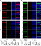
          Serine/threonine kinase 33 ( STK33), a member of the calcium/calmodulin‐dependent kinase ( CAMK), plays vital roles in a wide spectrum of cell processes. The present study was designed to investigate whether STK33 expressed in the mammalian cochlea and, if so, what effect STK33 exerted on aminoglycoside‐induced ototoxicity in House Ear Institute‐Organ of Corti 1 ( HEIOC1) cells. Immunofluorescence staining and western blotting were performed to investigate STK33 expression in cochlear hair cells ( HCs) and HEIOC1 cells with or without gentamicin treatment. CCK8, flow cytometry, immunofluorescence staining and western blotting were employed to detect the effects of STK33 knockdown, and/or U0126, and/or N‐acetyl‐L‐cysteine ( NAC) on the sensitivity to gentamicin‐induced ototoxicity in HEIOC1 cells. We found that STK33 was expressed in both mice cochlear HCs and HEIOC1 cells, and the expression of STK33 was significantly decreased in cochlear HCs and HEIOC1 cells after gentamicin exposure. STK33 knockdown resulted in an increase in the cleaved caspase‐3 and Bax expressions as well as cell apoptosis after gentamicin damage in HEIOC1 cells. Mechanistic studies revealed that knockdown of STK33 led to activated mitochondrial apoptosis pathway as well as augmented reactive oxygen species ( ROS) accumulation after gentamicin damage. Moreover, STK33 was involved in extracellular signal‐regulated kinase 1/2 pathway in primary culture of HCs and HEIOC1 cells in response to gentamicin insult. The findings from this work indicate that STK33 decreases the sensitivity to the apoptosis dependent on mitochondrial apoptotic pathway by regulating ROS generation after gentamicin treatment, which provides a new potential target for protection from the aminoglycoside‐induced ototoxicity.

          Related collections

          Most cited references34

          • Record: found
          • Abstract: found
          • Article: not found

          Mitogen-activated protein kinase: conservation of a three-kinase module from yeast to human.

          Mitogen-activated protein kinases (MAPK) are serine-threonine protein kinases that are activated by diverse stimuli ranging from cytokines, growth factors, neurotransmitters, hormones, cellular stress, and cell adherence. Mitogen-activated protein kinases are expressed in all eukaryotic cells. The basic assembly of MAPK pathways is a three-component module conserved from yeast to humans. The MAPK module includes three kinases that establish a sequential activation pathway comprising a MAPK kinase kinase (MKKK), MAPK kinase (MKK), and MAPK. Currently, there have been 14 MKKK, 7 MKK, and 12 MAPK identified in mammalian cells. The mammalian MAPK can be subdivided into five families: MAPKerk1/2, MAPKp38, MAPKjnk, MAPKerk3/4, and MAPKerk5. Each MAPK family has distinct biological functions. In Saccharomyces cerevisiae, there are five MAPK pathways involved in mating, cell wall remodelling, nutrient deprivation, and responses to stress stimuli such as osmolarity changes. Component members of the yeast pathways have conserved counterparts in mammalian cells. The number of different MKKK in MAPK modules allows for the diversity of inputs capable of activating MAPK pathways. In this review, we define all known MAPK module kinases from yeast to humans, what is known about their regulation, defined MAPK substrates, and the function of MAPK in cell physiology.
            Bookmark
            • Record: found
            • Abstract: found
            • Article: found
            Is Open Access

            Autophagy protects auditory hair cells against neomycin-induced damage

            ABSTRACT Aminoglycosides are toxic to sensory hair cells (HCs). Macroautophagy/autophagy is an essential and highly conserved self-digestion pathway that plays important roles in the maintenance of cellular function and viability under stress. However, the role of autophagy in aminoglycoside-induced HC injury is unknown. Here, we first found that autophagy activity was significantly increased, including enhanced autophagosome-lysosome fusion, in both cochlear HCs and HEI-OC-1 cells after neomycin or gentamicin injury, suggesting that autophagy might be correlated with aminoglycoside-induced cell death. We then used rapamycin, an autophagy activator, to increase the autophagy activity and found that the ROS levels, apoptosis, and cell death were significantly decreased after neomycin or gentamicin injury. In contrast, treatment with the autophagy inhibitor 3-methyladenine (3-MA) or knockdown of autophagy-related (ATG) proteins resulted in reduced autophagy activity and significantly increased ROS levels, apoptosis, and cell death after neomycin or gentamicin injury. Finally, after neomycin injury, the antioxidant N-acetylcysteine could successfully prevent the increased apoptosis and HC loss induced by 3-MA treatment or ATG knockdown, suggesting that autophagy protects against neomycin-induced HC damage by inhibiting oxidative stress. We also found that the dysfunctional mitochondria were not eliminated by selective autophagy (mitophagy) in HEI-OC-1 cells after neomycin treatment, suggesting that autophagy might not directly target the damaged mitochondria for degradation. This study demonstrates that moderate ROS levels can promote autophagy to recycle damaged cellular constituents and maintain cellular homeostasis, while the induction of autophagy can inhibit apoptosis and protect the HCs by suppressing ROS accumulation after aminoglycoside injury.
              Bookmark
              • Record: found
              • Abstract: found
              • Article: not found

              The aminoglycoside antibiotic dihydrostreptomycin rapidly enters mouse outer hair cells through the mechano-electrical transducer channels.

              The most serious side-effect of the widely used aminoglycoside antibiotics is irreversible intracellular damage to the auditory and vestibular hair cells of the inner ear. The mechanism of entry into the hair cells has not been unequivocally resolved. Here we report that extracellular dihydrostreptomycin not only blocks the mechano-electrical transducer channels of mouse outer hair cells at negative membrane potentials, as previously shown, but also enters the cells through these channels, which are located in the cells' mechanosensory hair bundles. The voltage-dependent blocking kinetics indicate an open-channel block mechanism, which can be well described by a two barrier-one binding site model, quantifying the antibiotic's block of the channel as well as its permeation in terms of the associated rate constants. The results identify the open transducer channels as the main route for aminoglycoside entry. Intracellularly applied dihydrostreptomycin also blocks the transducer channels, but at positive membrane potentials. However, the potency of the block was two orders of magnitude lower than that due to extracellular dihydrostreptomycin. Extracellular Ca2+ increases the free energy of the barrier nearest the extracellular side and of the binding site for dihydrostreptomycin. This reduces both the entry of dihydrostreptomycin into the channel and the channel's affinity for the drug. In vivo, where the extracellular Ca2+ concentration in the endolymph surrounding the hair bundles is < 100 microM, we predict that some 9000 dihydrostreptomycin molecules per second enter each hair cell at therapeutic drug concentrations.
                Bookmark

                Author and article information

                Contributors
                lijianfeng@hotmail.com
                wang.hb7585@hotmail.com
                Journal
                J Cell Mol Med
                J. Cell. Mol. Med
                10.1111/(ISSN)1582-4934
                JCMM
                Journal of Cellular and Molecular Medicine
                John Wiley and Sons Inc. (Hoboken )
                1582-1838
                1582-4934
                06 September 2018
                November 2018
                : 22
                : 11 ( doiID: 10.1111/jcmm.2018.22.issue-11 )
                : 5286-5299
                Affiliations
                [ 1 ] Otolaryngology‐Head and Neck Surgery Shandong Provincial Hospital Affiliated to Shandong University Jinan China
                [ 2 ] Shandong Provincial Key Laboratory of Otology Jinan China
                [ 3 ] Shandong Institute of Otolaryngology Jinan China
                Author notes
                [*] [* ] Correspondence Haibo Wang and Jianfeng Li, E‐mail: wang.hb7585@ 123456hotmail.com (HB) and lijianfeng@ 123456hotmail.com (JL)
                Author information
                http://orcid.org/0000-0003-3498-2690
                Article
                JCMM13792
                10.1111/jcmm.13792
                6201369
                30256516
                14184bec-1292-4887-b3ba-27185d9cd7cd
                © 2018 The Authors. Journal of Cellular and Molecular Medicine published by John Wiley & Sons Ltd and Foundation for Cellular and Molecular Medicine.

                This is an open access article under the terms of the http://creativecommons.org/licenses/by/4.0/ License, which permits use, distribution and reproduction in any medium, provided the original work is properly cited.

                History
                : 22 March 2018
                : 16 June 2018
                Page count
                Figures: 7, Tables: 0, Pages: 14, Words: 6820
                Funding
                Funded by: National 973 Basic Research Program of China
                Award ID: 2014CB541703
                Funded by: National Natural Science Foundation of China
                Award ID: 81072200
                Award ID: 81771008
                Categories
                Original Article
                Original Articles
                Custom metadata
                2.0
                jcmm13792
                November 2018
                Converter:WILEY_ML3GV2_TO_NLMPMC version:version=5.5.1 mode:remove_FC converted:25.10.2018

                Molecular medicine
                apoptosis,extracellular signal‐regulated kinase 1/2,gentamicin,reactive oxygen species,serine/threonine kinase 33

                Comments

                Comment on this article