49
views
0
recommends
+1 Recommend
0 collections
    0
    shares
      • Record: found
      • Abstract: found
      • Article: found
      Is Open Access

      High-Throughput Toxicity and Phenotypic Screening of 3D Human Neural Progenitor Cell Cultures on a Microarray Chip Platform

      research-article

      Read this article at

      Bookmark
          There is no author summary for this article yet. Authors can add summaries to their articles on ScienceOpen to make them more accessible to a non-specialist audience.

          Summary

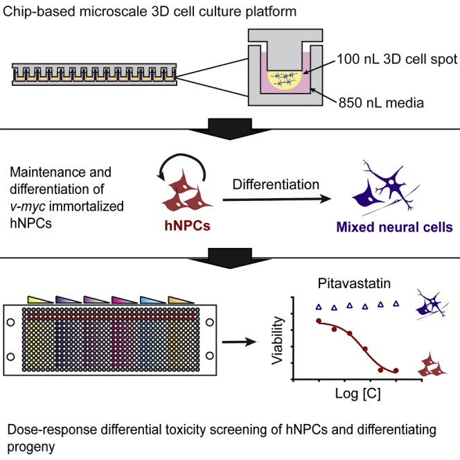
          A 3D cell culture chip was used for high-throughput screening of a human neural progenitor cell line. The differential toxicity of 24 compounds was determined on undifferentiated and differentiating NPCs. Five compounds led to significant differences in IC 50 values between undifferentiated and differentiating cultures. This platform has potential use in phenotypic screening to elucidate molecular toxicology on human stem cells.

          Graphical Abstract

          Highlights

          • Demonstrated chip platform for HTS of protein expression and toxicity of 3D cultures

          • Dose-response viability and proliferation of a 24-compound library on human NPC lines

          • Assessed differential toxicity between progenitors and differentiating progeny

          • Identified five compounds more toxic to undifferentiated progenitors

          Abstract

          Dordick and colleagues demonstrate a 3D cell culture chip for high-throughput screening of immortalized human neural progenitor cells. The dose-response toxicity of 24 compounds was determined on undifferentiated and differentiating hNPCs. Five compounds led to significant differences in IC 50 values between undifferentiated and differentiating cultures. This platform has potential use in phenotypic screening to elucidate molecular toxicology on human stem cells.

          Related collections

          Most cited references37

          • Record: found
          • Abstract: found
          • Article: not found

          In vitro differentiation of transplantable neural precursors from human embryonic stem cells.

          The remarkable developmental potential and replicative capacity of human embryonic stem (ES) cells promise an almost unlimited supply of specific cell types for transplantation therapies. Here we describe the in vitro differentiation, enrichment, and transplantation of neural precursor cells from human ES cells. Upon aggregation to embryoid bodies, differentiating ES cells formed large numbers of neural tube-like structures in the presence of fibroblast growth factor 2 (FGF-2). Neural precursors within these formations were isolated by selective enzymatic digestion and further purified on the basis of differential adhesion. Following withdrawal of FGF-2, they differentiated into neurons, astrocytes, and oligodendrocytes. After transplantation into the neonatal mouse brain, human ES cell-derived neural precursors were incorporated into a variety of brain regions, where they differentiated into both neurons and astrocytes. No teratoma formation was observed in the transplant recipients. These results depict human ES cells as a source of transplantable neural precursors for possible nervous system repair.
            Bookmark
            • Record: found
            • Abstract: found
            • Article: not found

            Migration of tumor cells in 3D matrices is governed by matrix stiffness along with cell-matrix adhesion and proteolysis.

            Cell migration on 2D surfaces is governed by a balance between counteracting tractile and adhesion forces. Although biochemical factors such as adhesion receptor and ligand concentration and binding, signaling through cell adhesion complexes, and cytoskeletal structure assembly/disassembly have been studied in detail in a 2D context, the critical biochemical and biophysical parameters that affect cell migration in 3D matrices have not been quantitatively investigated. We demonstrate that, in addition to adhesion and tractile forces, matrix stiffness is a key factor that influences cell movement in 3D. Cell migration assays in which Matrigel density, fibronectin concentration, and beta1 integrin binding are systematically varied show that at a specific Matrigel density the migration speed of DU-145 human prostate carcinoma cells is a balance between tractile and adhesion forces. However, when biochemical parameters such as matrix ligand and cell integrin receptor levels are held constant, maximal cell movement shifts to matrices exhibiting lesser stiffness. This behavior contradicts current 2D models but is predicted by a recent force-based computational model of cell movement in a 3D matrix. As expected, this 3D motility through an extracellular environment of pore size much smaller than cellular dimensions does depend on proteolytic activity as broad-spectrum matrix metalloproteinase (MMP) inhibitors limit the migration of DU-145 cells and also HT-1080 fibrosarcoma cells. Our experimental findings here represent, to our knowledge, discovery of a previously undescribed set of balances of cell and matrix properties that govern the ability of tumor cells to migration in 3D environments.
              Bookmark
              • Record: found
              • Abstract: found
              • Article: not found

              Clinical pharmacology of 5-fluorouracil.

              5-Fluorouracil, first introduced as a rationally synthesized anticancer agent 30 years ago, continues to be widely used in the management of several common malignancies including cancer of the colon, breast and skin. This drug, an analogue of the naturally occurring pyrimidine uracil, is metabolised via the same metabolic pathways as uracil. Although several potential sites of antitumour activity have been identified, the precise mechanism of action and the extent to which each of these sites contributes to tumour or host cell toxicity remains unclear. Several assay methods are available to quantify 5-fluorouracil in serum, plasma and other biological fluids. Unfortunately, there is no evidence that plasma drug concentrations can predict antitumour effect or host cell toxicity. The recent development of clinically useful pharmacodynamic assays provides an attractive alternative to plasma drug concentrations, since these assays allow the detection of active metabolites of 5-fluorouracil in biopsied tumour or normal tissue. 5-Fluorouracil is poorly absorbed after oral administration, with erratic bioavailability. The parenteral preparation is the major dosage form, used intravenously (bolus or continuous infusion). Recently, studies have demonstrated the pharmacokinetic rationale and clinical feasibility of hepatic arterial infusion and intraperitoneal administration of 5-fluorouracil. In addition, 5-fluorouracil continues to be used in topical preparations for the treatment of malignant skin cancers. Following parenteral administration of 5-fluorouracil, there is rapid distribution of the drug and rapid elimination with an apparent terminal half-life of approximately 8 to 20 minutes. The rapid elimination is primarily due to swift catabolism of the liver. As with all drugs, caution should be used in administering 5-fluorouracil in various pathophysiological states. In general, however, there are no set recommendations for dose adjustment in the presence of renal or hepatic dysfunction. Drug interactions continue to be described with other antineoplastic drugs, as well as with other classes of agents.
                Bookmark

                Author and article information

                Contributors
                Journal
                Stem Cell Reports
                Stem Cell Reports
                Stem Cell Reports
                Elsevier
                2213-6711
                27 October 2016
                08 November 2016
                27 October 2016
                : 7
                : 5
                : 970-982
                Affiliations
                [1 ]Chemical and Biological Engineering and Center for Biotechnology & Interdisciplinary Studies, Rensselaer Polytechnic Institute, Troy, NY 12180, USA
                [2 ]Chemical and Biomolecular Engineering, University of California, Berkeley, CA 94720, USA
                [3 ]Bioengineering, University of California, Berkeley, CA 94720, USA
                [4 ]Department of Bioengineering and Institute for Bioengineering and Biosciences, Instituto Superior Técnico, Universidade de Lisboa, Lisbon 1049-001, Portugal
                Author notes
                []Corresponding author dordick@ 123456rpi.edu
                Article
                S2213-6711(16)30220-X
                10.1016/j.stemcr.2016.10.001
                5106528
                11223422-ff41-46b8-a650-3e70e7efd8fc
                © 2016 The Author(s)

                This is an open access article under the CC BY license (http://creativecommons.org/licenses/by/4.0/).

                History
                : 9 May 2016
                : 30 September 2016
                : 3 October 2016
                Categories
                Article

                high-content screening,human neural stem cells,high-throughput microarray,screening,neurotoxicity,three-dimensional cell culture

                Comments

                Comment on this article