14
views
0
recommends
+1 Recommend
0 collections
    0
    shares
      • Record: found
      • Abstract: found
      • Article: found
      Is Open Access

      Ena/VASP clustering at microspike tips involves lamellipodin but not I-BAR proteins, and absolutely requires unconventional myosin-X

      research-article

      Read this article at

      Bookmark
          There is no author summary for this article yet. Authors can add summaries to their articles on ScienceOpen to make them more accessible to a non-specialist audience.

          Significance

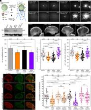
          The most prominent, protrusive structures of migrating cells on flat substrates include the sheet-like lamellipodia and actin-filament bundles termed filopodia or microspikes, the latter largely embedded in lamellipodia. Microspike formation requires clustering of Ena/VASP proteins at filament-barbed ends to enable sustained processive actin polymerization in the presence of heterodimeric capping protein. However, the factors and mechanisms mediating Ena/VASP clustering have remained elusive. Here, we systematically analyzed these processes in genetic knockout mutants derived from B16-F1 cells. Subsequent analysis revealed an unanticipated relationship of proteins implicated in control of dynamic actin protrusions showing, that contrary to previous assumptions, inverse BAR-domain proteins are not involved. Instead, we show that Ena/VASP clustering at microspike tips involves lamellipodin and strictly requires unconventional myosin-X.

          Abstract

          Sheet-like membrane protrusions at the leading edge, termed lamellipodia, drive 2D-cell migration using active actin polymerization. Microspikes comprise actin-filament bundles embedded within lamellipodia, but the molecular mechanisms driving their formation and their potential functional relevance have remained elusive. Microspike formation requires the specific activity of clustered Ena/VASP proteins at their tips to enable processive actin assembly in the presence of capping protein, but the factors and mechanisms mediating Ena/VASP clustering are poorly understood. Systematic analyses of B16-F1 melanoma mutants lacking potential candidate proteins revealed that neither inverse BAR-domain proteins, nor lamellipodin or Abi is essential for clustering, although they differentially contribute to lamellipodial VASP accumulation. In contrast, unconventional myosin-X (MyoX) identified here as proximal to VASP was obligatory for Ena/VASP clustering and microspike formation. Interestingly, and despite the invariable distribution of other relevant marker proteins, the width of lamellipodia in MyoX-KO mutants was significantly reduced as compared with B16-F1 control, suggesting that microspikes contribute to lamellipodium stability. Consistently, MyoX removal caused marked defects in protrusion and random 2D-cell migration. Strikingly, Ena/VASP-deficiency also uncoupled MyoX cluster dynamics from actin assembly in lamellipodia, establishing their tight functional association in microspike formation.

          Related collections

          Most cited references69

          • Record: found
          • Abstract: found
          • Article: not found

          Genome engineering using the CRISPR-Cas9 system.

          Targeted nucleases are powerful tools for mediating genome alteration with high precision. The RNA-guided Cas9 nuclease from the microbial clustered regularly interspaced short palindromic repeats (CRISPR) adaptive immune system can be used to facilitate efficient genome engineering in eukaryotic cells by simply specifying a 20-nt targeting sequence within its guide RNA. Here we describe a set of tools for Cas9-mediated genome editing via nonhomologous end joining (NHEJ) or homology-directed repair (HDR) in mammalian cells, as well as generation of modified cell lines for downstream functional studies. To minimize off-target cleavage, we further describe a double-nicking strategy using the Cas9 nickase mutant with paired guide RNAs. This protocol provides experimentally derived guidelines for the selection of target sites, evaluation of cleavage efficiency and analysis of off-target activity. Beginning with target design, gene modifications can be achieved within as little as 1-2 weeks, and modified clonal cell lines can be derived within 2-3 weeks.
            Bookmark
            • Record: found
            • Abstract: found
            • Article: found
            Is Open Access

            Easy quantitative assessment of genome editing by sequence trace decomposition

            The efficacy and the mutation spectrum of genome editing methods can vary substantially depending on the targeted sequence. A simple, quick assay to accurately characterize and quantify the induced mutations is therefore needed. Here we present TIDE, a method for this purpose that requires only a pair of PCR reactions and two standard capillary sequencing runs. The sequence traces are then analyzed by a specially developed decomposition algorithm that identifies the major induced mutations in the projected editing site and accurately determines their frequency in a cell population. This method is cost-effective and quick, and it provides much more detailed information than current enzyme-based assays. An interactive web tool for automated decomposition of the sequence traces is available. TIDE greatly facilitates the testing and rational design of genome editing strategies.
              Bookmark
              • Record: found
              • Abstract: found
              • Article: not found

              An improved smaller biotin ligase for BioID proximity labeling

              A smaller promiscuous biotin ligase for proximity biotinylation called BioID2 enables more-selective targeting of fusion proteins, requires less biotin supplementation, exhibits enhanced labeling of proximate proteins, and demonstrates the use of a flexible linker to modulate the biotin-labeling radius.
                Bookmark

                Author and article information

                Contributors
                Journal
                Proc Natl Acad Sci U S A
                Proc Natl Acad Sci U S A
                PNAS
                Proceedings of the National Academy of Sciences of the United States of America
                National Academy of Sciences
                0027-8424
                1091-6490
                4 January 2023
                10 January 2023
                4 January 2023
                : 120
                : 2
                : e2217437120
                Affiliations
                [1] aInstitute for Biophysical Chemistry, Hannover Medical School , 30625 Hannover, Germany
                [2] bIFOM ETS (Istituto Fondazione di Oncologia Molecolare ETS),  - The AIRC (Italian Association for Cancer Research) Institute of Molecular Oncology , 20139 Milan, Italy
                [3] cResearch Core Unit Proteomics, Hannover Medical School , 30625 Hannover, Germany
                [4] dDepartment of Oncology and Haemato-Oncology, University of Milan , 20139 Milan, Italy
                [5] eDivision of Molecular Cell Biology, Zoological Institute, Technische Universität Braunschweig , 38106 Braunschweig, Germany
                [6] fMolecular Cell Biology Group, Helmholtz Centre for Infection Research , 38124 Braunschweig, Germany
                Author notes
                2To whom correspondence may be addressed. Email: faix.jan@ 123456mh-hannover.de .

                Edited by Thomas Pollard, Department of Molecular Cellular and Developmental Biology, Yale University, New Haven, CT; received October 12, 2022; accepted November 14, 2022

                1T.P. and J.I.H. contributed equally to this work.

                Author information
                https://orcid.org/0000-0002-7222-8085
                https://orcid.org/0000-0002-1018-7679
                https://orcid.org/0000-0002-5077-9345
                https://orcid.org/0000-0001-7984-1889
                https://orcid.org/0000-0003-4244-4198
                https://orcid.org/0000-0003-1803-9192
                Article
                202217437
                10.1073/pnas.2217437120
                9926217
                36598940
                0f24f61d-6863-4b22-841e-a7c08999a274
                Copyright © 2023 the Author(s). Published by PNAS.

                This open access article is distributed under Creative Commons Attribution-NonCommercial-NoDerivatives License 4.0 (CC BY-NC-ND).

                History
                : 12 October 2022
                : 14 November 2022
                Page count
                Pages: 12, Words: 9029
                Funding
                Funded by: Deutsche Forschungsgemeinschaft (DFG), FundRef 501100001659;
                Award ID: FA330/9-2
                Award Recipient : Jan Faix
                Funded by: Deutsche Forschungsgemeinschaft (DFG), FundRef 501100001659;
                Award ID: FA330/13-1
                Award Recipient : Jan Faix
                Funded by: Associazione Italiana per la Ricerca sul Cancro (AIRC), FundRef 501100005010;
                Award ID: AIRC IG#18621
                Award Recipient : Giorgio Scita
                Funded by: Associazione Italiana per la Ricerca sul Cancro (AIRC), FundRef 501100005010;
                Award ID: 5Xmille#22759
                Award Recipient : Giorgio Scita
                Funded by: Italian Ministry of University and Scientific Research;
                Award ID: PRIN 2017 2017HWTP2K
                Award Recipient : Giorgio Scita Award Recipient : Klemens Rottner
                Funded by: Research Training Group;
                Award ID: GRK2223/1
                Award Recipient : Giorgio Scita Award Recipient : Klemens Rottner
                Categories
                video, Video
                research-article, Research Article
                cell-bio, Cell Biology
                409
                Biological Sciences
                Cell Biology

                microspikes,vasp clustering,i-bar proteins,lamellipodin,myosin-x

                Comments

                Comment on this article