23
views
0
recommends
+1 Recommend
0 collections
    0
    shares
      • Record: found
      • Abstract: found
      • Article: found
      Is Open Access

      GATA-dependent regulatory switches establish atrioventricular canal specificity during heart development

      research-article

      Read this article at

      Bookmark
          There is no author summary for this article yet. Authors can add summaries to their articles on ScienceOpen to make them more accessible to a non-specialist audience.

          Abstract

          The embryonic vertebrate heart tube develops an atrioventricular canal that divides the atrial and ventricular chambers, forms atrioventricular conduction tissue and organizes valve development. Here we assess the transcriptional mechanism underlying this localized differentiation process. We show that atrioventricular canal-specific enhancers are GATA-binding site-dependent and act as switches that repress gene activity in the chambers. We find that atrioventricular canal-specific gene loci are enriched in H3K27ac, a marker of active enhancers, in atrioventricular canal tissue and depleted in H3K27ac in chamber tissue. In the atrioventricular canal, Gata4 activates the enhancers in synergy with Bmp2/Smad signalling, leading to H3K27 acetylation. In contrast, in chambers, Gata4 cooperates with pan-cardiac Hdac1 and Hdac2 and chamber-specific Hey1 and Hey2, leading to H3K27 deacetylation and repression. We conclude that atrioventricular canal-specific enhancers are platforms integrating cardiac transcription factors, broadly active histone modification enzymes and localized co-factors to drive atrioventricular canal-specific gene activity.

          Abstract
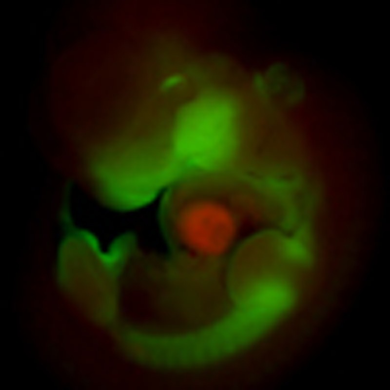
          The atrioventricular canal partitions the developing vertebrate heart. Here, the authors show that the cardiac transcription factor Gata4 together with histone modification enzymes and localized co-factors binds atrioventricular canal-specific enhancers, thereby repressing gene activity in the cardiac chambers.

          Related collections

          Most cited references55

          • Record: found
          • Abstract: found
          • Article: not found

          The incidence of congenital heart disease.

          This study was designed to determine the reasons for the variability of the incidence of congenital heart disease (CHD), estimate its true value and provide data about the incidence of specific major forms of CHD. The incidence of CHD in different studies varies from about 4/1,000 to 50/1,000 live births. The relative frequency of different major forms of CHD also differs greatly from study to study. In addition, another 20/1,000 live births have bicuspid aortic valves, isolated anomalous lobar pulmonary veins or a silent patent ductus arteriosus. The incidences reported in 62 studies published after 1955 were examined. Attention was paid to the ways in which the studies were conducted, with special reference to the increased use of echocardiography in the neonatal nursery. The total incidence of CHD was related to the relative frequency of ventricular septal defects (VSDs), the most common type of CHD. The incidences of individual major forms of CHD were determined from 44 studies. The incidence of CHD depends primarily on the number of small VSDs included in the series, and this number in turn depends upon how early the diagnosis is made. If major forms of CHD are stratified into trivial, moderate and severe categories, the variation in incidence depends mainly on the number of trivial lesions included. The incidence of moderate and severe forms of CHD is about 6/1,000 live births (19/1,000 live births if the potentially serious bicuspid aortic valve is included), and of all forms increases to 75/1,000 live births if tiny muscular VSDs present at birth and other trivial lesions are included. Given the causes of variation, there is no evidence for differences in incidence in different countries or times.
            Bookmark
            • Record: found
            • Abstract: found
            • Article: not found

            Histone deacetylases 1 and 2 redundantly regulate cardiac morphogenesis, growth, and contractility.

            Histone deacetylases (HDACs) tighten chromatin structure and repress gene expression through the removal of acetyl groups from histone tails. The class I HDACs, HDAC1 and HDAC2, are expressed ubiquitously, but their potential roles in tissue-specific gene expression and organogenesis have not been defined. To explore the functions of HDAC1 and HDAC2 in vivo, we generated mice with conditional null alleles of both genes. Whereas global deletion of HDAC1 results in death by embryonic day 9.5, mice lacking HDAC2 survive until the perinatal period, when they succumb to a spectrum of cardiac defects, including obliteration of the lumen of the right ventricle, excessive hyperplasia and apoptosis of cardiomyocytes, and bradycardia. Cardiac-specific deletion of either HDAC1 or HDAC2 does not evoke a phenotype, whereas cardiac-specific deletion of both genes results in neonatal lethality, accompanied by cardiac arrhythmias, dilated cardiomyopathy, and up-regulation of genes encoding skeletal muscle-specific contractile proteins and calcium channels. Our results reveal cell-autonomous and non-cell-autonomous functions for HDAC1 and HDAC2 in the control of myocardial growth, morphogenesis, and contractility, which reflect partially redundant roles of these enzymes in tissue-specific transcriptional repression.
              Bookmark
              • Record: found
              • Abstract: found
              • Article: not found

              Genetics of congenital heart disease: the glass half empty.

              Congenital heart disease (CHD) is the most common congenital anomaly in newborn babies. Cardiac malformations have been produced in multiple experimental animal models, by perturbing selected molecules that function in the developmental pathways involved in myocyte specification, differentiation, or cardiac morphogenesis. In contrast, the precise genetic, epigenetic, or environmental basis for these perturbations in humans remains poorly understood. Over the past few decades, researchers have tried to bridge this knowledge gap through conventional genome-wide analyses of rare Mendelian CHD families, and by sequencing candidate genes in CHD cohorts. Although yielding few, usually highly penetrant, disease gene mutations, these discoveries provided 3 notable insights. First, human CHD mutations impact a heterogeneous set of molecules that orchestrate cardiac development. Second, CHD mutations often alter gene/protein dosage. Third, identical pathogenic CHD mutations cause a variety of distinct malformations, implying that higher order interactions account for particular CHD phenotypes. The advent of contemporary genomic technologies including single nucleotide polymorphism arrays, next-generation sequencing, and copy number variant platforms are accelerating the discovery of genetic causes of CHD. Importantly, these approaches enable study of sporadic cases, the most common presentation of CHD. Emerging results from ongoing genomic efforts have validated earlier observations learned from the monogenic CHD families. In this review, we explore how continued use of these technologies and integration of systems biology is expected to expand our understanding of the genetic architecture of CHD.
                Bookmark

                Author and article information

                Journal
                Nat Commun
                Nat Commun
                Nature Communications
                Nature Pub. Group
                2041-1723
                28 April 2014
                : 5
                : 3680
                Affiliations
                [1 ]Department of Anatomy, Embryology and Physiology, Academic Medical Center, University of Amsterdam , Meibergdreef 15, 1105AZ Amsterdam, The Netherlands
                [2 ]Developmental Biochemistry, Biocenter and Comprehensive Heart Failure Center, University of Wuerzburg , Am Hubland, D-97074 Wuerzburg, Germany
                Author notes
                Article
                ncomms4680
                10.1038/ncomms4680
                4015328
                24770533
                0e848742-926c-4d29-8101-43e25e1e19d6
                Copyright © 2014, Nature Publishing Group, a division of Macmillan Publishers Limited. All Rights Reserved.

                This work is licensed under a Creative Commons Attribution-NonCommercial-NoDerivs 3.0 Unported License. The images or other third party material in this article are included in the article’s Creative Commons license, unless indicated otherwise in the credit line; if the material is not included under the Creative Commons license, users will need to obtain permission from the license holder to reproduce the material. To view a copy of this license, visit http://creativecommons.org/licenses/by-nc-nd/3.0/

                History
                : 12 November 2013
                : 17 March 2014
                Categories
                Article

                Uncategorized
                Uncategorized

                Comments

                Comment on this article