28
views
0
recommends
+1 Recommend
0 collections
    0
    shares
      • Record: found
      • Abstract: found
      • Article: found
      Is Open Access

      Haploid Germ Cells Generated in Organotypic Culture of Testicular Tissue From Prepubertal Boys

      research-article

      Read this article at

      Bookmark
          There is no author summary for this article yet. Authors can add summaries to their articles on ScienceOpen to make them more accessible to a non-specialist audience.

          Abstract

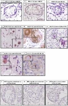
          While in mice various studies have described the completion of spermatogenesis in vitro using either organotypic culture of prepubertal testicular tissue or 3D culture of isolated cells, in humans it has not been possible to achieve germ cell differentiation from immature testicular tissue (ITT). In our study, we evaluated the ability of human ITT to differentiate via a long-term organotypic culture of frozen–thawed 1 mm 3 testicular fragments from five prepubertal boys in two different culture media. Tissue and supernatants were analyzed at regular intervals up to day 139. Sertoli cell (SC) viability and maturation was evaluated using immunohistochemistry (IHC) for SOX9, GDNF, anti-Mullerian hormone (AMH) and androgen receptor (AR), and AMH concentration in supernatants. Spermatogonia (SG) and proliferating cells were identified by MAGE-A4 (for SG) and Ki67 (for proliferating cells) via immunohistochemistry (IHC). Apoptotic cells were studied by active caspase 3. To evaluate Leydig cell (LC) functionality testosterone was measured in the supernatants and steroidogenic acute regulatory protein (STAR) IHC was performed. Germ cell differentiation was evaluated on Hematoxylin-Eosin histological sections, via IHC for synaptonemal complex 3 (SYCP3) for spermatocytes, Protein boule-like (BOLL) for spermatocytes and round spermatids, angiotensin-converting enzyme (ACE), protamine 2 and transition protein 1 (for elongated spermatids) and via chromogenic in situ hybridization (CISH). We reported the generation of meiotic and postmeiotic cells after 16 days of culture, as shown by the histological analyses, the presence of differentiation markers and the increase of haploid germ cells. We showed SC viability and maturation by a decrease of AMH secretion in the supernatants ( p ≤ 0.001) while the number of SOX9 positive cells did not show any variation. A decrease of spermatogonia ( p ≤ 0.001) was observed. The number of apoptotic cells did not vary. LC functionality was shown by the increase in STAR expression ( p ≤ 0.007) and a peak in testosterone secretion, followed by a reduction ( p ≤ 0.001) with stabilization. According to our knowledge, this is the first report of generation of haploid cells in human ITT. Differentiating germ cells have to be further evaluated for their ability to complete differentiation, their fecundability and epigenetic characteristics.

          Related collections

          Most cited references57

          • Record: found
          • Abstract: found
          • Article: not found

          In vitro production of functional sperm in cultured neonatal mouse testes.

          Spermatogenesis is one of the most complex and longest processes of sequential cell proliferation and differentiation in the body, taking more than a month from spermatogonial stem cells, through meiosis, to sperm formation. The whole process, therefore, has never been reproduced in vitro in mammals, nor in any other species with a very few exceptions in some particular types of fish. Here we show that neonatal mouse testes which contain only gonocytes or primitive spermatogonia as germ cells can produce spermatids and sperm in vitro with serum-free culture media. Spermatogenesis was maintained over 2 months in tissue fragments positioned at the gas-liquid interphase. The obtained spermatids and sperm resulted in healthy and reproductively competent offspring through microinsemination. In addition, neonatal testis tissues were cryopreserved and, after thawing, showed complete spermatogenesis in vitro. Our organ culture method could be applicable through further refinements to a variety of mammalian species, which will serve as a platform for future clinical application as well as mechanistic understanding of spermatogenesis.
            Bookmark
            • Record: found
            • Abstract: found
            • Article: not found

            A European perspective on testicular tissue cryopreservation for fertility preservation in prepubertal and adolescent boys.

            What clinical practices, patient management strategies and experimental methods are currently being used to preserve and restore the fertility of prepubertal boys and adolescent males?
              Bookmark
              • Record: found
              • Abstract: found
              • Article: not found

              Regulation of spermatogonial stem cell self-renewal and spermatocyte meiosis by Sertoli cell signaling.

              Spermatogenesis is a continuous and productive process supported by the self-renewal and differentiation of spermatogonial stem cells (SSCs), which arise from undifferentiated precursors known as gonocytes and are strictly controlled in a special 'niche' microenvironment in the seminiferous tubules. Sertoli cells, the only somatic cell type in the tubules, directly interact with SSCs to control their proliferation and differentiation through the secretion of specific factors. Spermatocyte meiosis is another key step of spermatogenesis, which is regulated by Sertoli cells on the luminal side of the blood-testis barrier through paracrine signaling. In this review, we mainly focus on the role of Sertoli cells in the regulation of SSC self-renewal and spermatocyte meiosis, with particular emphasis on paracrine and endocrine-mediated signaling pathways. Sertoli cell growth factors, such as glial cell line-derived neurotrophic factor (GDNF) and fibroblast growth factor 2 (FGF2), as well as Sertoli cell transcription factors, such as ETS variant 5 (ERM; also known as ETV5), nociceptin, neuregulin 1 (NRG1), and androgen receptor (AR), have been identified as the most important upstream factors that regulate SSC self-renewal and spermatocyte meiosis. Other transcription factors and signaling pathways (GDNF-RET-GFRA1 signaling, FGF2-MAP2K1 signaling, CXCL12-CXCR4 signaling, CCL9-CCR1 signaling, FSH-nociceptin/OPRL1, retinoic acid/FSH-NRG/ERBB4, and AR/RB-ARID4A/ARID4B) are also addressed.
                Bookmark

                Author and article information

                Contributors
                Journal
                Front Physiol
                Front Physiol
                Front. Physiol.
                Frontiers in Physiology
                Frontiers Media S.A.
                1664-042X
                09 October 2018
                2018
                : 9
                : 1413
                Affiliations
                [1] 1Department of Gynecology-Andrology, Cliniques Universitaires Saint-Luc , Brussels, Belgium
                [2] 2Institut de Recherche Expérimentale et Clinique (IREC), Université catholique de Louvain , Brussels, Belgium
                [3] 3Institut de Recherche Expérimentale et Clinique (IREC), Centre de Technologies Moléculaires Appliquées (CTMA) , Brussels, Belgium
                [4] 4Department of Clinical Biochemistry, Cliniques Universitaires Saint-Luc , Brussels, Belgium
                [5] 5Department of Anatomopathology, Cliniques Universitaires Saint-Luc , Brussels, Belgium
                Author notes

                Edited by: Richard Ivell, University of Nottingham, United Kingdom

                Reviewed by: Rod Mitchell, University of Edinburgh, United Kingdom; Séverine Mazaud-Guittot, Institut National de la Santé et de la Recherche Médicale (INSERM), France

                *Correspondence: Francesca de Michele, frademichele@ 123456gmail.com Christine Wyns, christine.wyns@ 123456uclouvain.be

                This article was submitted to Reproduction, a section of the journal Frontiers in Physiology

                Article
                10.3389/fphys.2018.01413
                6190924
                30356879
                0e15ed19-b25a-469e-a9a4-b5dd68aed8a7
                Copyright © 2018 de Michele, Poels, Vermeulen, Ambroise, Gruson, Guiot and Wyns.

                This is an open-access article distributed under the terms of the Creative Commons Attribution License (CC BY). The use, distribution or reproduction in other forums is permitted, provided the original author(s) and the copyright owner(s) are credited and that the original publication in this journal is cited, in accordance with accepted academic practice. No use, distribution or reproduction is permitted which does not comply with these terms.

                History
                : 02 July 2018
                : 18 September 2018
                Page count
                Figures: 12, Tables: 3, Equations: 0, References: 63, Pages: 18, Words: 0
                Categories
                Physiology
                Original Research

                Anatomy & Physiology
                prepubertal boys,testicular tissue cryopreservation,fertility preservation,in vitro maturation (ivm),haploid cells,human

                Comments

                Comment on this article