0
views
0
recommends
+1 Recommend
0 collections
    0
    shares
      • Record: found
      • Abstract: found
      • Article: found
      Is Open Access

      Role of Three-Dimensional Printing in Treatment Planning for Orthognathic Surgery: A Systematic Review

      review-article

      Read this article at

      Bookmark
          There is no author summary for this article yet. Authors can add summaries to their articles on ScienceOpen to make them more accessible to a non-specialist audience.

          Abstract

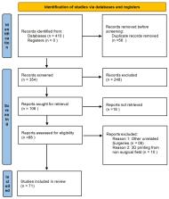
          Three-dimensional (3D) printing refers to a wide range of additive manufacturing processes that enable the construction of structures and models. It has been rapidly adopted for a variety of surgical applications, including the printing of patient-specific anatomical models, implants and prostheses, external fixators and splints, as well as surgical instrumentation and cutting guides. In comparison to traditional methods, 3D-printed models and surgical guides offer a deeper understanding of intricate maxillofacial structures and spatial relationships. This review article examines the utilization of 3D printing in orthognathic surgery, particularly in the context of treatment planning. It discusses how 3D printing has revolutionized this sector by providing enhanced visualization, precise surgical planning, reduction in operating time, and improved patient communication. Various databases, including PubMed, Google Scholar, ScienceDirect, and Medline, were searched with relevant keywords. A total of 410 articles were retrieved, of which 71 were included in this study. This article concludes that the utilization of 3D printing in the treatment planning of orthognathic surgery offers a wide range of advantages, such as increased patient satisfaction and improved functional and aesthetic outcomes.

          Related collections

          Most cited references92

          • Record: found
          • Abstract: found
          • Article: not found

          Surgical applications of three-dimensional printing: a review of the current literature & how to get started

          Three dimensional (3D) printing involves a number of additive manufacturing techniques that are used to build structures from the ground up. This technology has been adapted to a wide range of surgical applications at an impressive rate. It has been used to print patient-specific anatomic models, implants, prosthetics, external fixators, splints, surgical instrumentation, and surgical cutting guides. The profound utility of this technology in surgery explains the exponential growth. It is important to learn how 3D printing has been used in surgery and how to potentially apply this technology. PubMed was searched for studies that addressed the clinical application of 3D printing in all surgical fields, yielding 442 results. Data was manually extracted from the 168 included studies. We found an exponential increase in studies addressing surgical applications for 3D printing since 2011, with the largest growth in craniofacial, oromaxillofacial, and cardiothoracic specialties. The pertinent considerations for getting started with 3D printing were identified and are discussed, including, software, printing techniques, printing materials, sterilization of printing materials, and cost and time requirements. Also, the diverse and increasing applications of 3D printing were recorded and are discussed. There is large array of potential applications for 3D printing. Decreasing cost and increasing ease of use are making this technology more available. Incorporating 3D printing into a surgical practice can be a rewarding process that yields impressive results.
            Bookmark
            • Record: found
            • Abstract: found
            • Article: found
            Is Open Access

            From medical imaging data to 3D printed anatomical models

            Anatomical models are important training and teaching tools in the clinical environment and are routinely used in medical imaging research. Advances in segmentation algorithms and increased availability of three-dimensional (3D) printers have made it possible to create cost-efficient patient-specific models without expert knowledge. We introduce a general workflow that can be used to convert volumetric medical imaging data (as generated by Computer Tomography (CT)) to 3D printed physical models. This process is broken up into three steps: image segmentation, mesh refinement and 3D printing. To lower the barrier to entry and provide the best options when aiming to 3D print an anatomical model from medical images, we provide an overview of relevant free and open-source image segmentation tools as well as 3D printing technologies. We demonstrate the utility of this streamlined workflow by creating models of ribs, liver, and lung using a Fused Deposition Modelling 3D printer.
              Bookmark
              • Record: found
              • Abstract: found
              • Article: not found

              Accuracy of a computer-aided surgical simulation protocol for orthognathic surgery: a prospective multicenter study.

              The purpose of this prospective multicenter study was to assess the accuracy of a computer-aided surgical simulation (CASS) protocol for orthognathic surgery. The accuracy of the CASS protocol was assessed by comparing planned outcomes with postoperative outcomes of 65 consecutive patients enrolled from 3 centers. Computer-generated surgical splints were used for all patients. For the genioplasty, 1 center used computer-generated chin templates to reposition the chin segment only for patients with asymmetry. Standard intraoperative measurements were used without the chin templates for the remaining patients. The primary outcome measurements were the linear and angular differences for the maxilla, mandible, and chin when the planned and postoperative models were registered at the cranium. The secondary outcome measurements were the maxillary dental midline difference between the planned and postoperative positions and the linear and angular differences of the chin segment between the groups with and without the use of the template. The latter were measured when the planned and postoperative models were registered at the mandibular body. Statistical analyses were performed, and the accuracy was reported using root mean square deviation (RMSD) and the Bland-Altman method for assessing measurement agreement. In the primary outcome measurements, there was no statistically significant difference among the 3 centers for the maxilla and mandible. The largest RMSDs were 1.0 mm and 1.5° for the maxilla and 1.1 mm and 1.8° for the mandible. For the chin, there was a statistically significant difference between the groups with and without the use of the chin template. The chin template group showed excellent accuracy, with the largest positional RMSD of 1.0 mm and the largest orientation RMSD of 2.2°. However, larger variances were observed in the group not using the chin template. This was significant in the anteroposterior and superoinferior directions and the in pitch and yaw orientations. In the secondary outcome measurements, the RMSD of the maxillary dental midline positions was 0.9 mm. When registered at the body of the mandible, the linear and angular differences of the chin segment between the groups with and without the use of the chin template were consistent with the results found in the primary outcome measurements. Using this computer-aided surgical simulation protocol, the computerized plan can be transferred accurately and consistently to the patient to position the maxilla and mandible at the time of surgery. The computer-generated chin template provides greater accuracy in repositioning the chin segment than the intraoperative measurements. Copyright © 2013 American Association of Oral and Maxillofacial Surgeons. Published by Elsevier Inc. All rights reserved.
                Bookmark

                Author and article information

                Journal
                Cureus
                Cureus
                2168-8184
                Cureus
                Cureus (Palo Alto (CA) )
                2168-8184
                30 October 2023
                October 2023
                : 15
                : 10
                : e47979
                Affiliations
                [1 ] Oral and Maxillofacial Surgery, King Abdullah Medical City, Jeddah, SAU
                [2 ] Dentistry, Buraydah Colleges, Buraydah, SAU
                [3 ] General Dentistry, North of Riyadh Dental Clinic, Second Health Cluster, Riyadh, SAU
                [4 ] General Dentistry, Ibn Sina National College, Jeddah, SAU
                [5 ] Dentistry, Dar Al Uloom University, Riyadh, SAU
                [6 ] Vision College, King Abdulaziz University, Jeddah, SAU
                [7 ] Orthodontics, Ministry of Health, Riyadh, SAU
                [8 ] College of Dentistry, Imam Abdulrahman Bin Faisal University, Dammam, SAU
                [9 ] Oral and Maxillofacial Surgery, Ministry of Health, Jeddah, SAU
                [10 ] Maxillofacial Surgery, Ministry of Health, Dhahran, SAU
                Author notes
                Article
                10.7759/cureus.47979
                10686238
                38034130
                0d6989db-e7a3-49fb-9eb4-a4fd0c6f94df
                Copyright © 2023, Alhabshi et al.

                This is an open access article distributed under the terms of the Creative Commons Attribution License, which permits unrestricted use, distribution, and reproduction in any medium, provided the original author and source are credited.

                History
                : 30 October 2023
                Categories
                Dentistry

                lefort i osteotomy,bilateral lefort ii osteotomy,orthodontic surgery,digital dentistry,oral and maxillofacial surgeon

                Comments

                Comment on this article