28
views
0
recommends
+1 Recommend
0 collections
    0
    shares
      • Record: found
      • Abstract: found
      • Article: found
      Is Open Access

      Dynamic association of PfEMP1 and KAHRP in knobs mediates cytoadherence during Plasmodium invasion

      research-article

      Read this article at

      Bookmark
          There is no author summary for this article yet. Authors can add summaries to their articles on ScienceOpen to make them more accessible to a non-specialist audience.

          Abstract

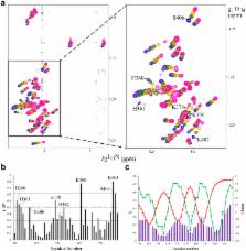
          Plasmodium falciparum infected erythrocytes display membrane knobs that are essential for their adherence to vascular endothelia and for prevention of clearance by the spleen. The knob associated histidine rich protein (KAHRP) is indispensable to knob formation and has been implicated in the recruitment and tethering of P. falciparum erythrocyte membrane protein–1 (PfEMP1) by binding to its cytoplasmic domain termed VARC. However, the precise mechanism of interaction between KAHRP and VARC is not very well understood. Here we report that both the proteins co-localize to membrane knobs of P. falciparum infected erythrocytes and have identified four positively charged linear sequence motifs of high intrinsic mobility on KAHRP that interact electrostatically with VARC in solution to form a fuzzy complex. The current study provides molecular insight into interaction between KAHRP and VARC in solution that takes place at membrane knobs.

          Related collections

          Most cited references43

          • Record: found
          • Abstract: found
          • Article: not found

          Protein NMR structure determination with automated NOE assignment using the new software CANDID and the torsion angle dynamics algorithm DYANA.

          Combined automated NOE assignment and structure determination module (CANDID) is a new software for efficient NMR structure determination of proteins by automated assignment of the NOESY spectra. CANDID uses an iterative approach with multiple cycles of NOE cross-peak assignment and protein structure calculation using the fast DYANA torsion angle dynamics algorithm, so that the result from each CANDID cycle consists of exhaustive, possibly ambiguous NOE cross-peak assignments in all available spectra and a three-dimensional protein structure represented by a bundle of conformers. The input for the first CANDID cycle consists of the amino acid sequence, the chemical shift list from the sequence-specific resonance assignment, and listings of the cross-peak positions and volumes in one or several two, three or four-dimensional NOESY spectra. The input for the second and subsequent CANDID cycles contains the three-dimensional protein structure from the previous cycle, in addition to the complete input used for the first cycle. CANDID includes two new elements that make it robust with respect to the presence of artifacts in the input data, i.e. network-anchoring and constraint-combination, which have a key role in de novo protein structure determinations for the successful generation of the correct polypeptide fold by the first CANDID cycle. Network-anchoring makes use of the fact that any network of correct NOE cross-peak assignments forms a self-consistent set; the initial, chemical shift-based assignments for each individual NOE cross-peak are therefore weighted by the extent to which they can be embedded into the network formed by all other NOE cross-peak assignments. Constraint-combination reduces the deleterious impact of artifact NOE upper distance constraints in the input for a protein structure calculation by combining the assignments for two or several peaks into a single upper limit distance constraint, which lowers the probability that the presence of an artifact peak will influence the outcome of the structure calculation. CANDID test calculations were performed with NMR data sets of four proteins for which high-quality structures had previously been solved by interactive protocols, and they yielded comparable results to these reference structure determinations with regard to both the residual constraint violations, and the precision and accuracy of the atomic coordinates. The CANDID approach has further been validated by de novo NMR structure determinations of four additional proteins. The experience gained in these calculations shows that once nearly complete sequence-specific resonance assignments are available, the automated CANDID approach results in greatly enhanced efficiency of the NOESY spectral analysis. The fact that the correct fold is obtained in cycle 1 of a de novo structure calculation is the single most important advance achieved with CANDID, when compared with previously proposed automated NOESY assignment methods that do not use network-anchoring and constraint-combination.
            Bookmark
            • Record: found
            • Abstract: found
            • Article: not found

            Cloning the P. falciparum gene encoding PfEMP1, a malarial variant antigen and adherence receptor on the surface of parasitized human erythrocytes.

            Plasmodium falciparum-infected human erythrocytes evade host immunity by expression of a cell-surface variant antigen and receptors for adherence to endothelial cells. These properties have been ascribed to P. falciparum erythrocyte membrane protein 1 (PfEMP1), an antigenically diverse malarial protein of 200-350 kDa on the surface of parasitized erythrocytes (PEs). We describe the cloning of two related PfEMP1 genes from the Malayan Camp (MC) parasite strain. Antibodies generated against recombinant protein fragments of the genes were specific for MC strain PfEMP1 protein. These antibodies reacted only with the surface of MC strain PEs and blocked adherence of these cells to CD36 but without effect on adherence to thrombospondin. Multiple forms of the PfEMP1 gene are apparent in MC parasites. The molecular basis for antigenic variation in malaria and adherence of infected erythrocytes to host cells can now be pursued.
              Bookmark
              • Record: found
              • Abstract: found
              • Article: not found

              The large diverse gene family var encodes proteins involved in cytoadherence and antigenic variation of Plasmodium falciparum-infected erythrocytes.

              The human malaria parasite Plasmodium falciparum evades host immunity by varying the antigenic and adhesive character of infected erythrocytes. We describe a large and extremely diverse family of P. falciparum genes (var) that encode 200-350 kDa proteins having the expected properties of antigenically variant adhesion molecules. Predicted amino acid sequences of var genes show a variable extracellular segment with domains having receptor-binding features, a transmembrane sequence, and a terminal segment that is a probable submembrane anchor. There are 50-150 var genes on multiple parasite chromosomes, and some are in clustered arrangements. var probes detect two classes of transcripts in steady-state RNA: 7-9 kb var transcripts, and an unusual family of 1.8-2.4 kb transcripts that may be involved in expression or rearrangements of var genes.
                Bookmark

                Author and article information

                Journal
                Sci Rep
                Sci Rep
                Scientific Reports
                Nature Publishing Group
                2045-2322
                02 March 2015
                2015
                : 5
                : 8617
                Affiliations
                [1 ]International Centre for Genetic Engineering and Biotechnology (ICGEB) , Aruna Asaf Ali Marg, New Delhi, India – 110 067
                [2 ]Department of Biosciences and Bioengineering, Indian Institute of Technology (IIT) , Bombay, Mumbai, India – 400 076
                Author notes
                Article
                srep08617
                10.1038/srep08617
                4345318
                25726759
                0d59176a-7f3c-46ac-8291-6770c5de54bd
                Copyright © 2015, Macmillan Publishers Limited. All rights reserved

                This work is licensed under a Creative Commons Attribution 4.0 International License. The images or other third party material in this article are included in the article's Creative Commons license, unless indicated otherwise in the credit line; if the material is not included under the Creative Commons license, users will need to obtain permission from the license holder in order to reproduce the material. To view a copy of this license, visit http://creativecommons.org/licenses/by/4.0/

                History
                : 14 September 2014
                : 26 January 2015
                Categories
                Article

                Uncategorized
                Uncategorized

                Comments

                Comment on this article