139
views
0
recommends
+1 Recommend
0 collections
    0
    shares
      • Record: found
      • Abstract: found
      • Article: found
      Is Open Access

      The molecular machinery of regulated cell death

      review-article

      Read this article at

      Bookmark
          There is no author summary for this article yet. Authors can add summaries to their articles on ScienceOpen to make them more accessible to a non-specialist audience.

          Abstract

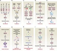
          Cells may die from accidental cell death (ACD) or regulated cell death (RCD). ACD is a biologically uncontrolled process, whereas RCD involves tightly structured signaling cascades and molecularly defined effector mechanisms. A growing number of novel non-apoptotic forms of RCD have been identified and are increasingly being implicated in various human pathologies. Here, we critically review the current state of the art regarding non-apoptotic types of RCD, including necroptosis, pyroptosis, ferroptosis, entotic cell death, netotic cell death, parthanatos, lysosome-dependent cell death, autophagy-dependent cell death, alkaliptosis and oxeiptosis. The in-depth comprehension of each of these lethal subroutines and their intercellular consequences may uncover novel therapeutic targets for the avoidance of pathogenic cell loss.

          Related collections

          Most cited references175

          • Record: found
          • Abstract: found
          • Article: not found

          ACSL4 dictates ferroptosis sensitivity by shaping cellular lipid composition.

          Ferroptosis is a form of regulated necrotic cell death controlled by glutathione peroxidase 4 (GPX4). At present, mechanisms that could predict sensitivity and/or resistance and that may be exploited to modulate ferroptosis are needed. We applied two independent approaches-a genome-wide CRISPR-based genetic screen and microarray analysis of ferroptosis-resistant cell lines-to uncover acyl-CoA synthetase long-chain family member 4 (ACSL4) as an essential component for ferroptosis execution. Specifically, Gpx4-Acsl4 double-knockout cells showed marked resistance to ferroptosis. Mechanistically, ACSL4 enriched cellular membranes with long polyunsaturated ω6 fatty acids. Moreover, ACSL4 was preferentially expressed in a panel of basal-like breast cancer cell lines and predicted their sensitivity to ferroptosis. Pharmacological targeting of ACSL4 with thiazolidinediones, a class of antidiabetic compound, ameliorated tissue demise in a mouse model of ferroptosis, suggesting that ACSL4 inhibition is a viable therapeutic approach to preventing ferroptosis-related diseases.
            Bookmark
            • Record: found
            • Abstract: found
            • Article: not found

            Ferroptosis: process and function.

            Ferroptosis is a recently recognized form of regulated cell death. It is characterized morphologically by the presence of smaller than normal mitochondria with condensed mitochondrial membrane densities, reduction or vanishing of mitochondria crista, and outer mitochondrial membrane rupture. It can be induced by experimental compounds (e.g., erastin, Ras-selective lethal small molecule 3, and buthionine sulfoximine) or clinical drugs (e.g., sulfasalazine, sorafenib, and artesunate) in cancer cells and certain normal cells (e.g., kidney tubule cells, neurons, fibroblasts, and T cells). Activation of mitochondrial voltage-dependent anion channels and mitogen-activated protein kinases, upregulation of endoplasmic reticulum stress, and inhibition of cystine/glutamate antiporter is involved in the induction of ferroptosis. This process is characterized by the accumulation of lipid peroxidation products and lethal reactive oxygen species (ROS) derived from iron metabolism and can be pharmacologically inhibited by iron chelators (e.g., deferoxamine and desferrioxamine mesylate) and lipid peroxidation inhibitors (e.g., ferrostatin, liproxstatin, and zileuton). Glutathione peroxidase 4, heat shock protein beta-1, and nuclear factor erythroid 2-related factor 2 function as negative regulators of ferroptosis by limiting ROS production and reducing cellular iron uptake, respectively. In contrast, NADPH oxidase and p53 (especially acetylation-defective mutant p53) act as positive regulators of ferroptosis by promotion of ROS production and inhibition of expression of SLC7A11 (a specific light-chain subunit of the cystine/glutamate antiporter), respectively. Misregulated ferroptosis has been implicated in multiple physiological and pathological processes, including cancer cell death, neurotoxicity, neurodegenerative diseases, acute renal failure, drug-induced hepatotoxicity, hepatic and heart ischemia/reperfusion injury, and T-cell immunity. In this review, we summarize the regulation mechanisms and signaling pathways of ferroptosis and discuss the role of ferroptosis in disease.
              Bookmark
              • Record: found
              • Abstract: not found
              • Article: not found

              Chemotherapy drugs induce pyroptosis through caspase-3 cleavage of a Gasdermin

                Bookmark

                Author and article information

                Contributors
                daolin.tang@utsouthwestern.edu
                kroemer@orange.fr
                Journal
                Cell Res
                Cell Res
                Cell Research
                Nature Publishing Group UK (London )
                1001-0602
                1748-7838
                4 April 2019
                4 April 2019
                May 2019
                : 29
                : 5
                : 347-364
                Affiliations
                [1 ]ISNI 0000 0000 8653 1072, GRID grid.410737.6, The Third Affiliated Hospital, Protein Modification and Degradation Lab, School of Basic Medical Sciences, , Guangzhou Medical University, ; 510510 Guangzhou, Guangdong China
                [2 ]ISNI 0000 0000 9482 7121, GRID grid.267313.2, Department of Surgery, , UT Southwestern Medical Center, ; Dallas, TX 75390 USA
                [3 ]ISNI 0000000104788040, GRID grid.11486.3a, Molecular Signaling and Cell Death Unit, VIB-UGent Center for Inflammation Research, , Flanders Institute for Biotechnology, ; 9052 Ghent, Belgium
                [4 ]ISNI 0000 0001 2069 7798, GRID grid.5342.0, Department for Biomedical Molecular Biology, , Ghent University, ; 9052 Ghent, Belgium
                [5 ]ISNI 0000 0001 0790 3681, GRID grid.5284.b, Laboratory of Pathophysiology, Faculty of Biomedical Sciences, , University of Antwerp, ; 2610 Wilrijk, Belgium
                [6 ]ISNI 0000 0001 2069 7798, GRID grid.5342.0, Methusalem program, , Ghent University, ; 9000 Ghent, Belgium
                [7 ]ISNI 0000 0001 2188 0914, GRID grid.10992.33, Université Paris Descartes, Sorbonne Paris Cité, ; 75006 Paris, France
                [8 ]GRID grid.417925.c, Equipe 11 labellisée Ligue Nationale contre le Cancer, Centre de Recherche des Cordeliers, ; 75006 Paris, France
                [9 ]ISNI 0000000121866389, GRID grid.7429.8, Institut National de la Santé et de la Recherche Médicale, ; U1138 Paris, France
                [10 ]ISNI 0000 0001 2308 1657, GRID grid.462844.8, Université Pierre et Marie Curie, ; 75006 Paris, France
                [11 ]ISNI 0000 0001 2284 9388, GRID grid.14925.3b, Metabolomics and Cell Biology Platforms, Gustave Roussy Cancer Campus, ; 94800 Villejuif, France
                [12 ]GRID grid.414093.b, Pôle de Biologie, Hôpital Européen Georges Pompidou, AP-HP, ; 75015 Paris, France
                [13 ]ISNI 0000 0000 9241 5705, GRID grid.24381.3c, Department of Women’s and Children’s Health, , Karolinska University Hospital, ; 17176 Stockholm, Sweden
                Author information
                http://orcid.org/0000-0002-6669-8822
                Article
                164
                10.1038/s41422-019-0164-5
                6796845
                30948788
                0be75cbe-45d9-4949-86c5-beb4c3d8bcd0
                © The Author(s) 2019

                Open Access This article is licensed under a Creative Commons Attribution 4.0 International License, which permits use, sharing, adaptation, distribution and reproduction in any medium or format, as long as you give appropriate credit to the original author(s) and the source, provide a link to the Creative Commons license, and indicate if changes were made. The images or other third party material in this article are included in the article’s Creative Commons license, unless indicated otherwise in a credit line to the material. If material is not included in the article’s Creative Commons license and your intended use is not permitted by statutory regulation or exceeds the permitted use, you will need to obtain permission directly from the copyright holder. To view a copy of this license, visit http://creativecommons.org/licenses/by/4.0/.

                History
                : 29 January 2019
                : 19 March 2019
                Categories
                Review Article
                Custom metadata
                © IBCB, SIBS, CAS 2019

                Cell biology
                cell signalling,cell death
                Cell biology
                cell signalling, cell death

                Comments

                Comment on this article