8
views
0
recommends
+1 Recommend
0 collections
    0
    shares
      • Record: found
      • Abstract: found
      • Article: not found

      Opening a Gateway for Chemiluminescence Cell Imaging: Distinctive Methodology for Design of Bright Chemiluminescent Dioxetane Probes

      research-article

      Read this article at

      ScienceOpenPublisherPMC
      Bookmark
          There is no author summary for this article yet. Authors can add summaries to their articles on ScienceOpen to make them more accessible to a non-specialist audience.

          Abstract

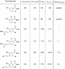
          Chemiluminescence probes are considered to be among the most sensitive diagnostic tools that provide high signal-to-noise ratio for various applications such as DNA detection and immunoassays. We have developed a new molecular methodology to design and foresee light-emission properties of turn-ON chemiluminescence dioxetane probes suitable for use under physiological conditions. The methodology is based on incorporation of a substituent on the benzoate species obtained during the chemiexcitation pathway of Schaap’s adamantylidene–dioxetane probe. The substituent effect was initially evaluated on the fluorescence emission generated by the benzoate species and then on the chemiluminescence of the dioxetane luminophores. A striking substituent effect on the chemiluminescence efficiency of the probes was obtained when acrylate and acrylonitrile electron-withdrawing groups were installed. The chemiluminescence quantum yield of the best probe was more than 3 orders of magnitude higher than that of a standard, commercially available adamantylidene–dioxetane probe. These are the most powerful chemiluminescence dioxetane probes synthesized to date that are suitable for use under aqueous conditions. One of our probes was capable of providing high-quality chemiluminescence cell images based on endogenous activity of β-galactosidase. This is the first demonstration of cell imaging achieved by a non-luciferin small-molecule probe with direct chemiluminescence mode of emission. We anticipate that the strategy presented here will lead to development of efficient chemiluminescence probes for various applications in the field of sensing and imaging.

          Abstract

          A new molecular methodology to design and foresee light-emission properties of turn-ON chemiluminescence dioxetane probes is presented. The probes are suitable for use under physiological conditions and thus could provide exceptional live cell images.

          Related collections

          Most cited references43

          • Record: found
          • Abstract: found
          • Article: not found

          Real-time imaging of oxidative and nitrosative stress in the liver of live animals for drug-toxicity testing

          Current drug-safety assays for hepatotoxicity rely on biomarkers with low predictive power. The production of radical species, specifically reactive oxygen species (ROS) and reactive nitrogen species (RNS), has been proposed as an early unifying event linking the bioactivation of drugs to hepatotoxicity and as a more direct and mechanistic indicator of hepatotoxic potential. Here we present a nanosensor for rapid, real-time in vivo imaging of drug-induced ROS and RNS for direct evaluation of acute hepatotoxicity. By combining fluorescence resonance energy transfer (FRET) and chemiluminescence resonance energy transfer (CRET), our semiconducting polymer–based nanosensor simultaneously and differentially detects RNS and ROS using two optically independent channels. Drug-induced hepatotoxicity and its remediation are imaged longitudinally in mice following systemic challenge with acetaminophen or isoniazid. Dose-dependent ROS and RNS activity is detected in the liver within minutes of drug challenge, preceding histological changes, protein nitration and DNA double strand break induction.
            Bookmark
            • Record: found
            • Abstract: found
            • Article: not found

            Bioluminescence imaging of myeloperoxidase activity in vivo.

            The myeloperoxidase (MPO) system of activated phagocytes is central to normal host defense mechanisms, and dysregulated MPO contributes to the pathogenesis of inflammatory disease states ranging from atherosclerosis to cancer. Here we show that upon systemic administration, the small molecule luminol enables noninvasive bioluminescence imaging (BLI) of MPO activity in vivo. Luminol-BLI allowed quantitative longitudinal monitoring of MPO activity in animal models of acute dermatitis, mixed allergic contact hypersensitivity, focal arthritis and spontaneous large granular lymphocytic tumors. Bioluminescence colocalized with histological sites of inflammation and was totally abolished in gene-deleted Mpo(-/-) mice, despite massive tissue infiltration of neutrophils and activated eosinophils, indicating that eosinophil peroxidase did not contribute to luminol-BLI in vivo. Thus, luminol-BLI provides a noninvasive, specific and highly sensitive optical readout of phagocyte-mediated MPO activity in vivo and may enable new diagnostic applications in a wide range of acute and chronic inflammatory conditions.
              Bookmark
              • Record: found
              • Abstract: found
              • Article: not found

              Intraparticle Energy Level Alignment of Semiconducting Polymer Nanoparticles to Amplify Chemiluminescence for Ultrasensitive In Vivo Imaging of Reactive Oxygen Species.

              Detection of reactive oxygen species (ROS), a hallmark of many pathological processes, is imperative to understanding, detection and treatment of many life-threatening diseases. However, methods capable of real-time in situ imaging of ROS in living animals are still very limited. We herein report the development and optimization of chemiluminescent semiconducting polymer nanoparticles (SPNs) for ultrasensitive in vivo imaging of hydrogen peroxide (H2O2). The chemiluminescence is amplified by adjusting the energy levels between the luminescence reporter and the chemiluminescence substrate to facilitate intermolecular electron transfer in the process of H2O2-activated luminescence. The optimized SPN can emit chemiluminescence with the quantum yield up to 2.30 × 10(-2) einsteins/mol and detect H2O2 down to 5 nM, which substantially outperforms the previous probes. Further doping of this SPN with a naphthalocyanine dye creates intraparticle chemiluminescence resonance energy transfer (CRET), leading to the near-infrared (NIR) luminescence responding to H2O2. By virtue of high brightness and ideal NIR optical window, SPN-NIR permits ultrasensitive imaging of H2O2 in the mouse models of peritonitis and neuroinflammation with the minute administration quantity. Thus, this study not only provides a category of optical probes that eliminates the need of external light excitation for imaging of H2O2, but also reveals the underlying principle to enhance the brightness of chemiluminescence systems.
                Bookmark

                Author and article information

                Journal
                ACS Cent Sci
                ACS Cent Sci
                oc
                acscii
                ACS Central Science
                American Chemical Society
                2374-7943
                2374-7951
                08 March 2017
                26 April 2017
                : 3
                : 4
                : 349-358
                Affiliations
                []School of Chemistry, Raymond and Beverly Sackler Faculty of Exact Sciences, Tel Aviv University , Tel Aviv 69978, Israel
                [§ ]Bioimaging Center, University of Geneva , Geneva, Switzerland
                Author notes
                [* ]Department of Organic Chemistry, School of Chemistry, Faculty of Exact Sciences, Tel Aviv University, Tel Aviv 69978, Israel. Tel: +972 (0) 3 640 8340. Fax: +972 (0) 3 640 9293. E-mail: chdoron@ 123456post.tau.ac.il .
                Article
                10.1021/acscentsci.7b00058
                5408346
                28470053
                0b3752e0-ee14-480f-b6fb-da04e3e9392e
                Copyright © 2017 American Chemical Society

                This is an open access article published under an ACS AuthorChoice License, which permits copying and redistribution of the article or any adaptations for non-commercial purposes.

                History
                : 01 February 2017
                Categories
                Research Article
                Custom metadata
                oc7b00058
                oc-2017-00058g

                Comments

                Comment on this article