50
views
0
recommends
+1 Recommend
1 collections
    0
    shares
      • Record: found
      • Abstract: found
      • Article: not found

      SERS Based Lateral Flow Immunoassay for Point-of-Care Detection of SARS-CoV-2 in Clinical Samples

      review-article

      Read this article at

      ScienceOpenPublisherPMC
      Bookmark
          There is no author summary for this article yet. Authors can add summaries to their articles on ScienceOpen to make them more accessible to a non-specialist audience.

          Abstract

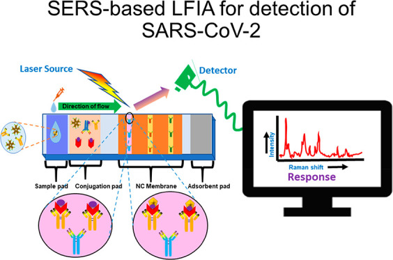
          The current scenario, an ongoing pandemic of COVID-19, places a dreadful burden on the healthcare system worldwide. Subsequently, there is a need for a rapid, user-friendly, and inexpensive on-site monitoring system for diagnosis. The early and rapid diagnosis of SARS-CoV-2 plays an important role in combating the outbreak. Although conventional methods such as PCR, RT-PCR, and ELISA, etc., offer a gold-standard solution to manage the pandemic, they cannot be implemented as a point-of-care (POC) testing arrangement. Moreover, surface-enhanced Raman spectroscopy (SERS) having a high enhancement factor provides quantitative results with high specificity, sensitivity, and multiplex detection ability but lacks in POC setup. In contrast, POC devices such as lateral flow immunoassay (LFIA) offer rapid, simple-to-use, cost-effective, reliable platform. However, LFIA has limitations in quantitative and sensitive analyses of SARS-CoV-2 detection. To resolve these concerns, herein we discuss a unique modality that is an integration of SERS with LFIA for quantitative analyses of SARS-CoV-2. The miniaturization ability of SERS-based devices makes them promising in biosensor application and has the potential to make a better alternative of conventional diagnostic methods. This review also demonstrates the commercially available and FDA/ICMR approved LFIA kits for on-site diagnosis of SARS-CoV-2.

          Related collections

          Most cited references149

          • Record: found
          • Abstract: found
          • Article: not found

          A familial cluster of pneumonia associated with the 2019 novel coronavirus indicating person-to-person transmission: a study of a family cluster

          Summary Background An ongoing outbreak of pneumonia associated with a novel coronavirus was reported in Wuhan city, Hubei province, China. Affected patients were geographically linked with a local wet market as a potential source. No data on person-to-person or nosocomial transmission have been published to date. Methods In this study, we report the epidemiological, clinical, laboratory, radiological, and microbiological findings of five patients in a family cluster who presented with unexplained pneumonia after returning to Shenzhen, Guangdong province, China, after a visit to Wuhan, and an additional family member who did not travel to Wuhan. Phylogenetic analysis of genetic sequences from these patients were done. Findings From Jan 10, 2020, we enrolled a family of six patients who travelled to Wuhan from Shenzhen between Dec 29, 2019 and Jan 4, 2020. Of six family members who travelled to Wuhan, five were identified as infected with the novel coronavirus. Additionally, one family member, who did not travel to Wuhan, became infected with the virus after several days of contact with four of the family members. None of the family members had contacts with Wuhan markets or animals, although two had visited a Wuhan hospital. Five family members (aged 36–66 years) presented with fever, upper or lower respiratory tract symptoms, or diarrhoea, or a combination of these 3–6 days after exposure. They presented to our hospital (The University of Hong Kong-Shenzhen Hospital, Shenzhen) 6–10 days after symptom onset. They and one asymptomatic child (aged 10 years) had radiological ground-glass lung opacities. Older patients (aged >60 years) had more systemic symptoms, extensive radiological ground-glass lung changes, lymphopenia, thrombocytopenia, and increased C-reactive protein and lactate dehydrogenase levels. The nasopharyngeal or throat swabs of these six patients were negative for known respiratory microbes by point-of-care multiplex RT-PCR, but five patients (four adults and the child) were RT-PCR positive for genes encoding the internal RNA-dependent RNA polymerase and surface Spike protein of this novel coronavirus, which were confirmed by Sanger sequencing. Phylogenetic analysis of these five patients' RT-PCR amplicons and two full genomes by next-generation sequencing showed that this is a novel coronavirus, which is closest to the bat severe acute respiatory syndrome (SARS)-related coronaviruses found in Chinese horseshoe bats. Interpretation Our findings are consistent with person-to-person transmission of this novel coronavirus in hospital and family settings, and the reports of infected travellers in other geographical regions. Funding The Shaw Foundation Hong Kong, Michael Seak-Kan Tong, Respiratory Viral Research Foundation Limited, Hui Ming, Hui Hoy and Chow Sin Lan Charity Fund Limited, Marina Man-Wai Lee, the Hong Kong Hainan Commercial Association South China Microbiology Research Fund, Sanming Project of Medicine (Shenzhen), and High Level-Hospital Program (Guangdong Health Commission).
            Bookmark
            • Record: found
            • Abstract: found
            • Article: not found

            SARS-CoV-2 Viral Load in Upper Respiratory Specimens of Infected Patients

            To the Editor: The 2019 novel coronavirus (SARS-CoV-2) epidemic, which was first reported in December 2019 in Wuhan, China, and has been declared a public health emergency of international concern by the World Health Organization, may progress to a pandemic associated with substantial morbidity and mortality. SARS-CoV-2 is genetically related to SARS-CoV, which caused a global epidemic with 8096 confirmed cases in more than 25 countries in 2002–2003. 1 The epidemic of SARS-CoV was successfully contained through public health interventions, including case detection and isolation. Transmission of SARS-CoV occurred mainly after days of illness 2 and was associated with modest viral loads in the respiratory tract early in the illness, with viral loads peaking approximately 10 days after symptom onset. 3 We monitored SARS-CoV-2 viral loads in upper respiratory specimens obtained from 18 patients (9 men and 9 women; median age, 59 years; range, 26 to 76) in Zhuhai, Guangdong, China, including 4 patients with secondary infections (1 of whom never had symptoms) within two family clusters (Table S1 in the Supplementary Appendix, available with the full text of this letter at NEJM.org). The patient who never had symptoms was a close contact of a patient with a known case and was therefore monitored. A total of 72 nasal swabs (sampled from the mid-turbinate and nasopharynx) (Figure 1A) and 72 throat swabs (Figure 1B) were analyzed, with 1 to 9 sequential samples obtained from each patient. Polyester flock swabs were used for all the patients. From January 7 through January 26, 2020, a total of 14 patients who had recently returned from Wuhan and had fever (≥37.3°C) received a diagnosis of Covid-19 (the illness caused by SARS-CoV-2) by means of reverse-transcriptase–polymerase-chain-reaction assay with primers and probes targeting the N and Orf1b genes of SARS-CoV-2; the assay was developed by the Chinese Center for Disease Control and Prevention. Samples were tested at the Guangdong Provincial Center for Disease Control and Prevention. Thirteen of 14 patients with imported cases had evidence of pneumonia on computed tomography (CT). None of them had visited the Huanan Seafood Wholesale Market in Wuhan within 14 days before symptom onset. Patients E, I, and P required admission to intensive care units, whereas the others had mild-to-moderate illness. Secondary infections were detected in close contacts of Patients E, I, and P. Patient E worked in Wuhan and visited his wife (Patient L), mother (Patient D), and a friend (Patient Z) in Zhuhai on January 17. Symptoms developed in Patients L and D on January 20 and January 22, respectively, with viral RNA detected in their nasal and throat swabs soon after symptom onset. Patient Z reported no clinical symptoms, but his nasal swabs (cycle threshold [Ct] values, 22 to 28) and throat swabs (Ct values, 30 to 32) tested positive on days 7, 10, and 11 after contact. A CT scan of Patient Z that was obtained on February 6 was unremarkable. Patients I and P lived in Wuhan and visited their daughter (Patient H) in Zhuhai on January 11 when their symptoms first developed. Fever developed in Patient H on January 17, with viral RNA detected in nasal and throat swabs on day 1 after symptom onset. We analyzed the viral load in nasal and throat swabs obtained from the 17 symptomatic patients in relation to day of onset of any symptoms (Figure 1C). Higher viral loads (inversely related to Ct value) were detected soon after symptom onset, with higher viral loads detected in the nose than in the throat. Our analysis suggests that the viral nucleic acid shedding pattern of patients infected with SARS-CoV-2 resembles that of patients with influenza 4 and appears different from that seen in patients infected with SARS-CoV. 3 The viral load that was detected in the asymptomatic patient was similar to that in the symptomatic patients, which suggests the transmission potential of asymptomatic or minimally symptomatic patients. These findings are in concordance with reports that transmission may occur early in the course of infection 5 and suggest that case detection and isolation may require strategies different from those required for the control of SARS-CoV. How SARS-CoV-2 viral load correlates with culturable virus needs to be determined. Identification of patients with few or no symptoms and with modest levels of detectable viral RNA in the oropharynx for at least 5 days suggests that we need better data to determine transmission dynamics and inform our screening practices.
              Bookmark
              • Record: found
              • Abstract: found
              • Article: not found

              Antibody responses to SARS-CoV-2 in patients with COVID-19

              We report acute antibody responses to SARS-CoV-2 in 285 patients with COVID-19. Within 19 days after symptom onset, 100% of patients tested positive for antiviral immunoglobulin-G (IgG). Seroconversion for IgG and IgM occurred simultaneously or sequentially. Both IgG and IgM titers plateaued within 6 days after seroconversion. Serological testing may be helpful for the diagnosis of suspected patients with negative RT-PCR results and for the identification of asymptomatic infections.
                Bookmark

                Author and article information

                Journal
                ACS Appl Bio Mater
                ACS Appl Bio Mater
                mt
                aabmcb
                ACS Applied Bio Materials
                American Chemical Society
                2576-6422
                17 March 2021
                : acsabm.1c00102
                Affiliations
                []Microfluidics & MEMS Centre, CSIR−Advanced Materials and Processes Research Institute (AMPRI) , Hoshangabad Road, Bhopal 462026, India
                []Academy of Scientific and Innovative Research (AcSIR) , Ghaziabad 201002, India
                Author notes
                Article
                10.1021/acsabm.1c00102
                7986978
                0ae698c3-85bb-401c-810d-80852013726b
                © 2021 American Chemical Society

                This article is made available via the PMC Open Access Subset for unrestricted RESEARCH re-use and analyses in any form or by any means with acknowledgement of the original source. These permissions are granted for the duration of the World Health Organization (WHO) declaration of COVID-19 as a global pandemic.

                History
                : 27 January 2021
                : 04 March 2021
                Categories
                Review
                Custom metadata
                mt1c00102
                mt1c00102

                surface-enhanced raman spectroscopy,lateral flow immunoassay,sars-cov-2,covid-19,poc devices

                Comments

                Comment on this article