13
views
0
recommends
+1 Recommend
0 collections
    0
    shares
      • Record: found
      • Abstract: found
      • Article: found
      Is Open Access

      Papulonecrotic Tuberculid

      research-article

      Read this article at

      Bookmark
          There is no author summary for this article yet. Authors can add summaries to their articles on ScienceOpen to make them more accessible to a non-specialist audience.

          Abstract

          A 30-year-old man presented with a 6 year history of recurrent, multiple asymptomatic raised lesions over his back and bilateral upper limbs. He had been treated repeatedly for a case of recurrent boils with oral and topical antibiotics. Some of the lesions had healed spontaneously leaving behind unsightly scars. The patient denied any history of associated fever, chronic cough, weight loss, and drug intake prior to the onset of lesions. There was no recognized contact with tuberculosis patients. General and systemic examination was essentially normal. Dermatological examination revealed the presence of multiple, well-defined, hyperpigmented crusted papules of 0.5–1.0 cm in size, distributed symmetrically over his entire back, extensor surface of bilateral forearm, arm, and bilateral dorsum of foot, interspersed with atrophic varioliform scarring (Figures 1 and 2). Routine laboratory workup was normal. Tuberculin (Mantoux) was strongly positive at 72 hours (23 × 23 mm) with central necrosis (Figure 3). Sputum for acid fast bacilli culture, chest radiograph, ultrasound abdomen, and pelvis did 
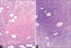
not reveal any abnormality. A biopsy specimen taken from a crusted papule over his forearm (Figure 4) showed fibrinoid necrosis, surrounded by mixed inflammatory infiltrate filling the entire dermis along with a few ill-defined epitheloid granulomas and lymphocytoclastic vasculitis. These are consistent with papulonecrotic tuberculid. Figure 1. Multiple, hyperpigmented, crusted papules with varioliform scarring seen over the back. Figure 2. Atrophic varioliform scarring over extensor aspect of left forearm.
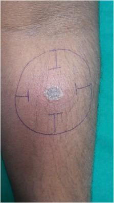
 Figure 3. Positive Mantoux reaction (23 × 23).
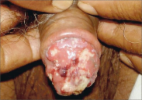
 Figure 4. Single, crusted papule from which skin biopsy was taken. The patient was started on a four drug combination therapy of rifampicin, isoniazid, pyrazinamide, and ethambutol for two months initially followed by a combination of rifampicin and isoniazid to complete a total of 6 months of standard antitubercular therapy. The patient responded well and all of the lesions eventually healed. Tuberculids are hypersensitivity reactions to Mycobacterium tuberculosis or its products in individuals with good immunity. 1 Papulonecrotic tuberculid is a relatively uncommon manifestation of cutaneous tuberculosis. 2 It presents as chronic, recurrent, symmetrical eruption of necrotizing papules that ulcerate, crust, and heal after a few weeks with varioliform scarring. 3 Hallmarks of this disease include positive mantoux test, evidence of present or past tuberculosis, inability to isolate M. tuberculosis in the skin lesions, resolution of lesions with atrophic varioliform lesions, and response to antitubercular therapy. 1 In a country like India where the prevalence of tuberculosis is high, existence of papulonecrotic tuberculid is possible and the focus of the infection may not be demonstrable in the majority of cases. Once diagnosed patients respond very well to antituberculosis therapy. It is important to consider papulonecrotic tuberculid as a differential diagnosis in any patient with recurrent eruption that heals with scarring in an endemic area.

          Related collections

          Most cited references3

          • Record: found
          • Abstract: found
          • Article: found
          Is Open Access

          PENILE PAPULONECROTIC TUBERCULID: REVISITED

          Sir, Papulonecrotic tuberculid causing penile ulcers is extremely rare.1 Herewith, we report a case of papulonecrotic tuberculide of the penis in a 56-years-old male. A 56-year-old married male patient was referred to our department with multiple asymptomatic non-healing ulcers over the glans penis of one month duration. He was a heterosexual individual and his wife did not have any genital lesions or discharge. The patient denied any history of pre-marital and extra-marital sexual contact. On physical examination, there were multiple, superficial and deep tender ulcers on the glans penis with ragged, irregular margins and floor covered with necrotic yellow slough (Fig. 1). The urethral meatus was hidden by these ulcerative lesions. Rest of the genital examination was normal. There was no inguinal lymphadenopathy. His systemic examination was unremarkable. Fig. 1 Glans penis showing multiple ulcers The hemogram revealed elevated erythrocyte sedimentation rate (50 mm in the first hour). Tuberculin (Mantoux) test was strongly positive (20 mm × 20 mm). Gram stain of the discharge from the ulcers demonstrated pus cells, Gram positive cocci, and Gram negative bacilli and discharge from the ulcers grew Staphylococcus aureus, Escherichia coli, and Enterococcus faecalis. Ziehl Neelsen stain of the pus did not demonstrate any acid-fast bacilli (AFB). Tzanck smear from ulcer was negative for multinucleated giant cells. Urine sediment examination for AFB and urine culture were noncontributory. Radiological and ultrasound evaluation of the genitourinary system was normal. HIV antibodies test and VDRL test were nonreactive. Systemic evaluation for any focus of tuberculosis was unremarkable. Biopsy from the edge of the ulcer (glans penis) revealed ulcerated epidermis. In the deep dermis, by the side of ulceration, there were caseating tuberculous granulomas along with perivascular infiltrate with vessel wall thickening and endothelial cell swelling. Fite stain for AFB was negative. These features were consistent with papulonecrotic tuberculide. AFB culture of biopsy specimen was negative. Repeated courses of antibiotic therapy did not yield desired results; hence, antitubercular therapy was initiated keeping in mind the possibility of papuloneurotic tuberculide of the penis. Four-drug combination therapy of rifampicin, isoniazid, pyrazinamide, and ethambutol was given for initial 2 months followed by combination of rifampicin and isoniazid to complete total 6 months of standard antitubercular therapy. The lesions started responding to therapy in next two weeks and complete healing with residual depressed scars occurred after three months of therapy (Fig. 2). Fig. 2 Healing with residual depressed scars after 3 months of therapy Tuberculosis of the penis is rare, even in third world countries where the prevalence of tuberculosis remains relatively high.1 Till 1999, only 161 cases of penile tuberculosis were reported.2 Understandably, papulonecrotic tuberculide involving the glans penis is even rarer.3 4 Tuberculides are hypersensitivity reactions to Mycobacterium tuberculosis or its products in individuals with good immunity.5 These cases are characterized by positive tuberculin test, evidence of present or past tuberculosis, absence of M. tuberculosis in the skin lesions and response to antitubercular treatment.5 However, a focus of tuberculosis elsewhere in the body may not be demonstrable in majority of the cases with papulonecrotic tuberculide as in our case.2 6 Papulonecrotic tuberculides are characterized by recurrent eruptions of asymptomatic, dusky red papules, which ulcerate and crust, and heal after a few weeks with varioliform scarring.5 7 These occur symmetrically and predominantly on the extensor aspects (legs, knees, elbows, hands and feet) of the extremities. Other areas that may be rarely affected by papulonecrotic tuberculides are the ears, face, buttocks, perniotic areas and penis.5 In Japan, penile tuberculide has been considered a disease entity.4 Thus, it is important to remember tuberculosis as an underlying cause of penile ulcers, more so in countries like India where prevalence of tuberculosis is still high.
            Bookmark
            • Record: found
            • Abstract: found
            • Article: not found

            Papulonecrotic tuberculid: report of a clinical case

            Papulonecrotic tuberculid is a rare, chronic, recurrent and symmetric eruption of necrotizing skin papules arising in crops, involving primarily the limbs. It occurs in less than 5% of active tuberculosis. Typically bacilli are not detected in any of the cutaneous lesions. Extracutaneous tuberculosis primary focus is only detected in 30–40% of the cases, being the lymph nodes the most common location. Antituberculosis treatment is rapidly effective and is often the main confirmation of the diagnosis.
              Bookmark
              • Record: found
              • Abstract: found
              • Article: found
              Is Open Access

              Papulonecrotic Tuberculid with Positive Acid-fast Bacilli

              Two patients suffered from ‘boils’ on their arms and/or thighs for several months. A diagnosis of papulonecrotic tuberculid (PNT) was made based on clinical, laboratory parameters, histopathology, and a prompt response to multi-drug anti-tuberculosis treatment. We checked the pathological sections carefully and finally found a small amount of positive acid-fast bacilli. We analyzed the clinical and histopathological features of PNT in order to offer reference of preventing and controlling the disease.
                Bookmark

                Author and article information

                Journal
                Am J Trop Med Hyg
                Am. J. Trop. Med. Hyg
                tpmd
                tropmed
                The American Journal of Tropical Medicine and Hygiene
                The American Society of Tropical Medicine and Hygiene
                0002-9637
                1476-1645
                11 October 2017
                11 October 2017
                : 97
                : 4
                : 987-988
                Affiliations
                [1]Department of Dermatology, Venereology and Leprosy, Sri Manakula Vinayagar Medical College and Hospital, Puducherry, India
                Author notes
                [* ]Address correspondence to Valeti Meghana, Department of Dermatology, Venereology and Leprosy, Sri Manakula Vinayagar Medical College and Hospital, Kalitheerthalkuppam, Puducherry 605107, India. E-mail: sweety.magi6@ 123456gmail.com

                Authors’ addresses: Valeti Meghana and Gowtham Saravanan, Department of Dermatology, Sri Manakula Vinayagar Medical College and Hospital, Pondicherry, India, E-mails: sweety.magi6@ 123456gmail.com and gowthamms2010@ 123456gmail.com . Kaliaperumal Karthikeyan, Department of Dermatology, Venereology and Leprosy, Sri Manakula Vinayagar Medical College and Hospital, Puducherry, India, E-mail: karthikderm@ 123456gmail.com .

                Article
                tpmd170377
                10.4269/ajtmh.17-0377
                5637623
                29031284
                096e5673-df42-4e85-a464-a1dfdab3dfbc
                © The American Society of Tropical Medicine and Hygiene

                This is an open-access article distributed under the terms of the Creative Commons Attribution License, which permits unrestricted use, distribution, and reproduction in any medium, provided the original author and source are credited.

                History
                : 12 May 2017
                : 24 May 2017
                Page count
                Pages: 2
                Categories
                Images in Clinical Tropical Medicine

                Infectious disease & Microbiology
                Infectious disease & Microbiology

                Comments

                Comment on this article