665
views
0
recommends
+1 Recommend
0 collections
    12
    shares
      • Record: found
      • Abstract: found
      • Article: found
      Is Open Access

      Bilateral osseous stenosis of the internal auditory canal: case report Translated title: A proposito di un caso di stenosi bilaterale ossea del condotto uditivo interno

      case-report

      Read this article at

      Bookmark
          There is no author summary for this article yet. Authors can add summaries to their articles on ScienceOpen to make them more accessible to a non-specialist audience.

          SUMMARY

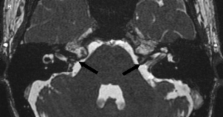
          Osteomas as well as exostoses of the internal auditory canal are rare, benign, usually slow-growing lesions. The most common localizations of these temporal bone lesions are the mastoid cortex and the external auditory canal. A rare case is reported of bilateral osseous stenosis of the internal auditory canal, in the absence of clinical (auditory, vestibular and facial nerve) symptoms. In the absence of auditory, vestibular and/or facial nerve symptoms, long-term follow-up should be assessed; surgical intervention may be warranted only if symptoms are present.

          RIASSUNTO

          Gli osteomi e le esostosi del condotto uditivo interno rappresentano una condizione patologica di raro riscontro, benigna e di solito caratterizzata da un lento accrescimento. Le localizzazioni più frequenti di tali lesioni dell'osso temporale sono la corticale mastoidea e il condotto uditivo esterno. Gli autori riportano un raro caso di stenosi bilaterale ossea del condotto uditivo interno, in assenza di sintomi o segni audiologici. In assenza di sintomatologia uditiva, vestibolare o deficit del faciale, è necessario sottoporre il paziente a follow-up a lungo termine, mentre l'opzione chirurgica è da considerarsi solo qualora vi siano manifestazioni cliniche importanti.

          Related collections

          Most cited references22

          • Record: found
          • Abstract: not found
          • Book: not found

          Pathology of the ear

            Bookmark
            • Record: found
            • Abstract: not found
            • Article: not found

            Osteopetrosis (Marble bone disease) of the temporal bone.

            H Hamersma (1970)
              Bookmark
              • Record: found
              • Abstract: found
              • Article: not found

              Osteoma of the internal auditory canal.

              We present a case of an osteoma of the internal auditory canal, which was surgically removed with symptomatic improvement. We describe CT, MRI, histological and operative findings, with a review of the proposed aetiology. The implications for imaging in the investigation of retrocochlear lesions are discussed.
                Bookmark

                Author and article information

                Journal
                Acta Otorhinolaryngol Ital
                Pacini
                Acta Otorhinolaryngologica Italica
                Pacini Editore SpA
                0392-100X
                1827-675X
                June 2011
                29 December 2010
                : 31
                : 3
                : 177-180
                Affiliations
                Audiology Department
                [1 ] Neuroradiology Department, University Hospital of Ferrara, Italy
                Author notes
                Address for correspondence: Dr. A. Ciorba, U.O. di Audiologia, Università di Ferrara, c.so Giovecca 203, 44100 Ferrara, Italy. Fax: +39 0532 236887. E-mail: andrea.ciorba@ 123456unife.it
                Article
                Pacini
                3185815
                22064765
                06e09e87-5c49-4e48-be25-eddb14095aff
                © Copyright by Società Italiana di Otorinolaringologia e Chirurgia Cervico-Facciale

                This is an Open Access article distributed under the terms of the Creative Commons Attribution Non-Commercial No Derivatives License, which permits for noncommercial use, distribution, and reproduction in any digital medium, provided the original work is properly cited and is not altered in any way. For details, please refer to http://creativecommons.org/licenses/by-nc-nd/3.0/

                History
                : 15 March 2010
                : 21 June 2010
                Categories
                Case Report

                Otolaryngology
                skull base,imaging,exostosis,internal auditory canal,osteomas
                Otolaryngology
                skull base, imaging, exostosis, internal auditory canal, osteomas

                Comments

                Comment on this article