44
views
0
recommends
+1 Recommend
0 collections
    0
    shares
      • Record: found
      • Abstract: found
      • Article: found
      Is Open Access

      Evaluation of Internal Auditory Canal Structures in Tinnitus of Unknown Origin

      research-article

      Read this article at

      Bookmark
          There is no author summary for this article yet. Authors can add summaries to their articles on ScienceOpen to make them more accessible to a non-specialist audience.

          Abstract

          Objectives

          The aim of the present study was to evaluate the internal auditory canal (IAC) and the nerves inside it to define possible structural differences in cases with subjective tinnitus of unknown origin.

          Methods

          Cases applying to the ear, nose and throat department with the complaint of tinnitus with unknown origin and having normal physical examination and test results were included in the study (n=78). Patients admitted to the radiology clinic for routine cranial magnetic resonance imaging (MRI) and whose MRI findings revealed no pathologies were enrolled as the control group (n=79). Data for the control group were obtained from the radiology department and informed consent was obtained from all the patients. Diameters of the IAC and the nerves inside it were measured through enhanced images obtained by routine temporal bone MRIs in all cases. Statistical evaluations were performed using Student t-test and statistical significance was defined as P<0.05.

          Results

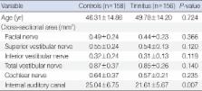
          Measurements of IAC diameters revealed statistically significant differences between the controls and the tinnitus group ( P<0.05). Regarding the diameters of the cochlear nerve, facial nerve, inferior vestibular nerve, superior vestibular nerve, and total vestibular nerve, no statistically significant difference was found between the controls and the tinnitus group.

          Conclusion

          Narrowed IAC has to be assessed as an etiological factor in cases with subjective tinnitus of unknown origin.

          Related collections

          Most cited references26

          • Record: found
          • Abstract: found
          • Article: not found

          Quantitative evaluation of convolution-based methods for medical image interpolation.

          Interpolation is required in a variety of medical image processing applications. Although many interpolation techniques are known from the literature, evaluations of these techniques for the specific task of applying geometrical transformations to medical images are still lacking. In this paper we present such an evaluation. We consider convolution-based interpolation methods and rigid transformations (rotations and translations). A large number of sinc-approximating kernels are evaluated, including piecewise polynomial kernels and a large number of windowed sinc kernels, with spatial supports ranging from two to ten grid intervals. In the evaluation we use images from a wide variety of medical image modalities. The results show that spline interpolation is to be preferred over all other methods, both for its accuracy and its relatively low computational cost.
            Bookmark
            • Record: found
            • Abstract: found
            • Article: not found

            Internal auditory canal morphology in children with cochlear nerve deficiency.

            To describe the internal auditory canal (IAC) and inner ear morphologic characteristics of children with cochlear nerve (CN) deficiency. Retrospective case series. Tertiary referral center. Fourteen children with small or absent (deficient) CNs have been identified by means of high-resolution magnetic resonance imaging (MRI). MRI of the brain. Clinical evaluation. Review of medical records, audiological testing results, and imaging studies. Images were evaluated for the structure of the cochlear, vestibular and facial nerves, IACs and inner ears. Audiometric thresholds were evaluated in all subjects. Fourteen children with small or absent (deficient) CNs have been identified by means of high-resolution MRI. A review of the medical records, audiologic testing results, and imaging studies was undertaken. The images were evaluated for the structure of the cochlear, vestibular and facial nerves, IACs, and inner ears. The audiometric thresholds were evaluated in all subjects. Among the 14 patients, 5 had known syndromes. MRI allowed an exact specification of the nervous structures within all ears with normal-size IACs. Precise characterization of the nerves in ears with small IACs was more difficult, requiring a consideration of both imaging findings and functional parameters. Five children had bilateral deficient CNs, whereas the remaining 9 subjects were affected unilaterally. Thus, 19 ears had CN deficiency (absent CN, 16; small CN, 3). Eleven ears had normal-size IACs and deficient CNs. Of the 9 ears with small IACs, 8 had deficient CNs (absent, 7; small, 1) on the basis of both MRI and functional assessments. Two ears with small IACs had clear morphologic and/or functional evidence for the presence of a CN: one had a small-size CN on MRI, whereas another had a single nerve in a small IAC with present facial and auditory functions. The findings of this study suggest that CN deficiency is not an uncommon cause of congenital hearing loss. The findings that most ears with CN deficiency had normal IAC morphology and that two ears with small IACs had CNs present indicate that IAC morphology is an unreliable surrogate marker of CN integrity. On the basis of these findings, we think that high-resolution MRI, rather than CT imaging, should be performed in all cases of pediatric hearing loss, especially in those cases where profound hearing loss has been documented. For ears with small IACs, the resolution of MRI currently remains limiting. In these cases, the determination of CN status frequently requires a variety of anatomic (CT and MRI) and functional tests (auditory brainstem response, otoacoustic emissions, behavioral audiometry, and physical examination).
              Bookmark
              • Record: found
              • Abstract: found
              • Article: not found

              Appearance of normal cranial nerves on steady-state free precession MR images.

              As radiologic imaging technology improves and more intricate details of the anatomy can be evaluated, images provide more precise diagnostic information and allow better localization of abnormalities. For example, standard T2-weighted magnetic resonance (MR) imaging sequences adequately depicted only the larger cranial nerves, whereas current steady-state free precession (SSFP) sequences are capable of depicting the cisternal segments of all 12 cranial nerves. SSFP sequences provide submillimetric spatial resolution and high contrast resolution between cerebrospinal fluid and solid structures, allowing the reconstruction of elegant multiplanar images that highlight the course of each nerve. These sequences have become a mainstay in the evaluation of the cerebellopontine angles and inner ear. Usually referred to by their trade names or acronyms (eg, constructive interference steady state, or CISS, and fast imaging employing steady-state acquisition, or FIESTA), SSFP sequences allow precise differentiation between branches of the facial and vestibulocochlear nerves, accurate detection of small masses in the cerebellopontine angles and internal auditory canals, and detailed evaluation of endolymph and perilymph within the inner ear. To take full advantage of these imaging sequences, radiologists must be familiar with the appearances of similar anatomic details of all 12 cranial nerves on SSFP MR images. Copyright RSNA, 2009
                Bookmark

                Author and article information

                Journal
                Clin Exp Otorhinolaryngol
                Clin Exp Otorhinolaryngol
                CEO
                Clinical and Experimental Otorhinolaryngology
                Korean Society of Otorhinolaryngology-Head and Neck Surgery
                1976-8710
                2005-0720
                September 2014
                01 August 2014
                : 7
                : 3
                : 160-164
                Affiliations
                [1 ]Department of Otolaryngology, Elazig Training and Research Hospital, Elazig, Turkey.
                [2 ]Department of Radiology, Elazig Training and Research Hospital, Elazig, Turkey.
                [3 ]Department of Computer Engineering, Faculty of Engineering, Firat University, Elazig, Turkey.
                Author notes
                Corresponding author: Cahit Polat. Department of Otolaryngology, Elazig Training and Research Hospital, 23300 Elazig, Turkey. Tel: +90-424-238-1000, Fax: +90-424-212-1461, drcahitpolat@ 123456yahoo.com
                Article
                10.3342/ceo.2014.7.3.160
                4135149
                25177429
                0525e015-5b14-469f-bc74-4d8aeddf3587
                Copyright © 2014 by Korean Society of Otorhinolaryngology-Head and Neck Surgery.

                This is an open-access article distributed under the terms of the Creative Commons Attribution Non-Commercial License ( http://creativecommons.org/licenses/by-nc/3.0/) which permits unrestricted non-commercial use, distribution, and reproduction in any medium, provided the original work is properly cited.

                History
                : 11 March 2013
                : 11 April 2013
                : 22 April 2013
                Categories
                Original Article

                Otolaryngology
                tinnitus,diameter of internal auditory canal,cochlear nerve,vestibular nerve,facial nerve,magnetic resonance imaging

                Comments

                Comment on this article