49
views
0
recommends
+1 Recommend
0 collections
    0
    shares
      • Record: found
      • Abstract: found
      • Article: found
      Is Open Access

      Florid cemento-osseous dysplasia: Report of a case documented with clinical, radiographic, biochemical and histological findings

      case-report

      Read this article at

      Bookmark
          There is no author summary for this article yet. Authors can add summaries to their articles on ScienceOpen to make them more accessible to a non-specialist audience.

          Abstract

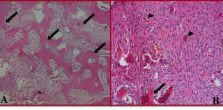
          Florid cemento-osseous dysplasia (FCOD) has been described as a condition that characteristically affects the jaws of middle-aged black women. This condition has also been classified as gigantiform cementoma, chronic sclerosing osteomyelitis, sclerosing osteitis, multiple estenosis and sclerotic cemental masses. It usually exhibits as multiple radiopaque cementum-like masses distributed throughout the jaws. Radiographically, FCOD appears as dense, lobulated masses, often symmetrically located in various regions of the jaws. Computed tomography, because of its ability to give axial, sagittal, and frontal views, is useful in the evaluation of these lesions. This article reports the case of a 45-year-old white man who was diagnosed with FCOD on the basis of clinical, radiographic, biochemical and histological findings. It is of major importance to realize that all dentists have a unique opportunity as well as ethical obligation to assist in the struggle against wrong dental treatments that might save patients dental health. This case report illustrates the point that periapical radiolucencies may represent benign fibro-osseous lesions that may be overlooked or result in unnecessary endodontic treatment.

          Key words:Florid cemento-osseous dysplasia, florid osseous dysplasia, fibro-osseous lesions.

          Related collections

          Most cited references14

          • Record: found
          • Abstract: found
          • Article: not found

          Fibro-osseous lesions of the jaws.

          During the 15 years since the author's 1970 paper on fibro-osseous lesions of the jaws, the subject has continued to interest the clinician and pathologist. Several new entities have been delineated during this period, and new concepts and controversies regarding the diagnosis and management of these lesions have arisen. A classification of fibro-osseous jaw lesions that the author has found to be workable is presented, and criteria for their diagnosis and management based on the authors' experience is discussed.
            Bookmark
            • Record: found
            • Abstract: found
            • Article: not found

            Florid osseous dysplasia. A clinical-pathologic study of thirty-four cases.

            Thirty-four patients with florid osseous dysplasia were studied. The majority were asymptomatic Negro women: Seventeen biopsy-proven simple bone cysts were found in affected quadrants of fourteen patients. Radiographs displayed a spectrum of sclerotic and ground-glass opacities limited to alveolar processes but not to root apices. Biopsy material was studied in all cases, and biochemical analyses of serum and cyst fluid were performed on some patients. Test results and skeletal radiographs indicate that the disease is limited to the jaws. Patients have remained asymptomatic with little alteration of radiographic patterns. Three cysts failed to heal following treatment, others filled with radiographically abnormal tissue. Chronic osteomyelitis may infrequently complicate the disease. These cases appear to represent the most exuberant manifestation of this reactive fibro-osseous jaw disease.
              Bookmark
              • Record: found
              • Abstract: found
              • Article: not found

              Florid cemento-osseous dysplasia: Report of a case documented with computed tomography and 3D imaging.

              Florid cemento-osseous dysplasia is an uncommon disease of the jaws. This article reports the case of a 49-year-old black woman who was diagnosed with florid cemento-osseous dysplasia on the basis of conventional radiographic findings. Computed tomography and three-dimensional imaging of the mandible were performed, and they enabled detailed localization of the bone defects. The role of computed tomography in the evaluation of cemento-osseous lesions is discussed.
                Bookmark

                Author and article information

                Journal
                J Clin Exp Dent
                J Clin Exp Dent
                Medicina Oral S.L.
                Journal of Clinical and Experimental Dentistry
                Medicina Oral S.L.
                1989-5488
                1 February 2013
                February 2013
                : 5
                : 1
                : e58-e61
                Affiliations
                [1 ]Department of Oral and Maxillofacial Surgery, Faculty of Dentistry, Gazi University, Ankara, Turkey
                [2 ]Department of Prosthodontics, Faculty of Dentistry, Gazi University, Ankara, Turkey
                [3 ]Department of Oral Pathology, Faculty of Dentistry, Gazi University, Ankara, Turkey
                Author notes
                Yukar?bahçelievler Mah Eski 24. Sk. 4/6 , 06490 Ankara, Turkey , E-mail: harikakutluay@ 123456yahoo.com

                Conflict of interest statement: The authors have declared that no conflict of interest exist.

                Article
                50854
                10.4317/jced.50854
                3892229
                24455053
                0609f0a0-673b-4514-9fc9-a28535c79177
                Copyright: © 2013 Medicina Oral S.L.

                This is an open-access article distributed under the terms of the Creative Commons Attribution License, which permits unrestricted use, distribution, and reproduction in any medium, provided the original work is properly cited.

                History
                : 18 October 2012
                : 16 April 2012
                Categories
                Case Report
                Oral Medicine and Pathology

                Comments

                Comment on this article