11
views
0
recommends
+1 Recommend
0 collections
    0
    shares
      • Record: found
      • Abstract: not found
      • Article: not found

      Brain-wide glymphatic enhancement and clearance in humans assessed with MRI

      Read this article at

      ScienceOpenPublisherPMC
      Bookmark
          There is no author summary for this article yet. Authors can add summaries to their articles on ScienceOpen to make them more accessible to a non-specialist audience.

          Abstract

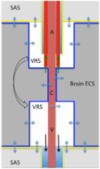
          <p class="first" id="d8414236e234">To what extent does the subarachnoid cerebrospinal fluid (CSF) compartment communicate directly with the extravascular compartment of human brain tissue? Interconnection between the subarachnoid CSF compartment and brain perivascular spaces is reported in some animal studies, but with controversy, and in vivo CSF tracer studies in humans are lacking. In the present work, we examined the distribution of a CSF tracer in the human brain by MRI over a prolonged time span. For this, we included a reference cohort, representing close to healthy individuals, and a cohort of patients with dementia and anticipated compromise of CSF circulation (idiopathic normal pressure hydrocephalus). The MRI contrast agent gadobutrol, which is confined to the extravascular brain compartment by the intact blood-brain barrier, was used as a CSF tracer. Standardized T1-weighted MRI scans were performed before and after intrathecal gadobutrol at defined time points, including at 24 hours, 48 hours, and 4 weeks. All MRI scans were aligned and brain regions were segmented using FreeSurfer, and changes in normalized T1 signals over time were quantified as percentage change from baseline. The study provides in vivo evidence of access to all human brain subregions of a substance administered intrathecally. Clearance of the tracer substance was delayed in the dementia cohort. These observations translate previous findings in animal studies into humans and open new prospects concerning intrathecal treatment regimens, extravascular contrast-enhanced MRI, and assessment of brain clearance function. </p><p class="first" id="d8414236e237">This study provides evidence for brain-wide communication with the subarachnoid CSF space in humans and reduced glymphatic clearance in a cohort with iNPH dementia. </p>

          Related collections

          Most cited references41

          • Record: found
          • Abstract: found
          • Article: not found

          A hybrid approach to the skull stripping problem in MRI.

          We present a novel skull-stripping algorithm based on a hybrid approach that combines watershed algorithms and deformable surface models. Our method takes advantage of the robustness of the former as well as the surface information available to the latter. The algorithm first localizes a single white matter voxel in a T1-weighted MRI image, and uses it to create a global minimum in the white matter before applying a watershed algorithm with a preflooding height. The watershed algorithm builds an initial estimate of the brain volume based on the three-dimensional connectivity of the white matter. This first step is robust, and performs well in the presence of intensity nonuniformities and noise, but may erode parts of the cortex that abut bright nonbrain structures such as the eye sockets, or may remove parts of the cerebellum. To correct these inaccuracies, a surface deformation process fits a smooth surface to the masked volume, allowing the incorporation of geometric constraints into the skull-stripping procedure. A statistical atlas, generated from a set of accurately segmented brains, is used to validate and potentially correct the segmentation, and the MRI intensity values are locally re-estimated at the boundary of the brain. Finally, a high-resolution surface deformation is performed that accurately matches the outer boundary of the brain, resulting in a robust and automated procedure. Studies by our group and others outperform other publicly available skull-stripping tools. Copyright 2004 Elsevier Inc.
            Bookmark
            • Record: found
            • Abstract: found
            • Article: not found

            Brain-wide pathway for waste clearance captured by contrast-enhanced MRI.

            The glymphatic system is a recently defined brain-wide paravascular pathway for cerebrospinal fluid (CSF) and interstitial fluid (ISF) exchange that facilitates efficient clearance of solutes and waste from the brain. CSF enters the brain along para-arterial channels to exchange with ISF, which is in turn cleared from the brain along para-venous pathways. Because soluble amyloid β clearance depends on glymphatic pathway function, we proposed that failure of this clearance system contributes to amyloid plaque deposition and Alzheimer's disease progression. Here we provide proof of concept that glymphatic pathway function can be measured using a clinically relevant imaging technique. Dynamic contrast-enhanced MRI was used to visualize CSF-ISF exchange across the rat brain following intrathecal paramagnetic contrast agent administration. Key features of glymphatic pathway function were confirmed, including visualization of para-arterial CSF influx and molecular size-dependent CSF-ISF exchange. Whole-brain imaging allowed the identification of two key influx nodes at the pituitary and pineal gland recesses, while dynamic MRI permitted the definition of simple kinetic parameters to characterize glymphatic CSF-ISF exchange and solute clearance from the brain. We propose that this MRI approach may provide the basis for a wholly new strategy to evaluate Alzheimer's disease susceptibility and progression in the live human brain.
              Bookmark
              • Record: found
              • Abstract: found
              • Article: found
              Is Open Access

              A new look at cerebrospinal fluid circulation

              According to the traditional understanding of cerebrospinal fluid (CSF) physiology, the majority of CSF is produced by the choroid plexus, circulates through the ventricles, the cisterns, and the subarachnoid space to be absorbed into the blood by the arachnoid villi. This review surveys key developments leading to the traditional concept. Challenging this concept are novel insights utilizing molecular and cellular biology as well as neuroimaging, which indicate that CSF physiology may be much more complex than previously believed. The CSF circulation comprises not only a directed flow of CSF, but in addition a pulsatile to and fro movement throughout the entire brain with local fluid exchange between blood, interstitial fluid, and CSF. Astrocytes, aquaporins, and other membrane transporters are key elements in brain water and CSF homeostasis. A continuous bidirectional fluid exchange at the blood brain barrier produces flow rates, which exceed the choroidal CSF production rate by far. The CSF circulation around blood vessels penetrating from the subarachnoid space into the Virchow Robin spaces provides both a drainage pathway for the clearance of waste molecules from the brain and a site for the interaction of the systemic immune system with that of the brain. Important physiological functions, for example the regeneration of the brain during sleep, may depend on CSF circulation.
                Bookmark

                Author and article information

                Journal
                JCI Insight
                American Society for Clinical Investigation
                2379-3708
                July 12 2018
                July 12 2018
                July 12 2018
                July 12 2018
                : 3
                : 13
                Article
                10.1172/jci.insight.121537
                6124518
                29997300
                06070509-5d9b-4a44-8045-9cc8e5966c9e
                © 2018
                History

                Comments

                Comment on this article