33
views
0
recommends
+1 Recommend
0 collections
    0
    shares
      • Record: found
      • Abstract: found
      • Article: found
      Is Open Access

      Intracellular calcium movements during excitation–contraction coupling in mammalian slow-twitch and fast-twitch muscle fibers

      research-article

      Read this article at

      Bookmark
          There is no author summary for this article yet. Authors can add summaries to their articles on ScienceOpen to make them more accessible to a non-specialist audience.

          Abstract

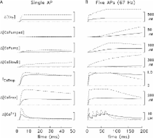
          In skeletal muscle fibers, action potentials elicit contractions by releasing calcium ions (Ca 2+) from the sarcoplasmic reticulum. Experiments on individual mouse muscle fibers micro-injected with a rapidly responding fluorescent Ca 2+ indicator dye reveal that the amount of Ca 2+ released is three- to fourfold larger in fast-twitch fibers than in slow-twitch fibers, and the proportion of the released Ca 2+ that binds to troponin to activate contraction is substantially smaller.

          Related collections

          Most cited references82

          • Record: found
          • Abstract: found
          • Article: not found

          Involvement of dihydropyridine receptors in excitation-contraction coupling in skeletal muscle.

          The transduction of action potential to muscle contraction (E-C coupling) is an example of fast communication between plasma membrane events and the release of calcium from an internal store, which in muscle is the sarcoplasmic reticulum (SR). One theory is that the release channels of the SR are controlled by voltage-sensing molecules or complexes, located in the transverse tubular (T)-membrane, which produce, as membrane voltage varies, 'intramembrane charge movements', but nothing is known about the structure of such sensors. Receptors of the Ca-channel-blocking dihydropyridines present in many tissues, are most abundant in T-tubular muscle fractions from which they can be isolated as proteins. Fewer than 5% of muscle dihydropyridines are functional Ca channels; there is no known role for the remainder in skeletal muscle physiology. We report here that low concentrations of a dihydropyridine inhibit charge movements and SR calcium release in parallel. The effect has a dependence on membrane voltage analogous to that of specific binding of dihydropyridines. We propose specifically that the molecule that generates charge movement is the dihydropyridine receptor.
            Bookmark
            • Record: found
            • Abstract: not found
            • Article: not found

            Voltage dependent charge movement of skeletal muscle: a possible step in excitation-contraction coupling.

              Bookmark
              • Record: found
              • Abstract: found
              • Article: not found

              Restoration of excitation-contraction coupling and slow calcium current in dysgenic muscle by dihydropyridine receptor complementary DNA.

              Microinjection of an expression plasmid that carries complementary DNA encoding the receptor for dihydropyridine calcium channel blockers of skeletal muscle restores both excitation-contraction coupling and slow calcium current in cultured skeletal muscle cells from mice with muscular dysgenesis. This suggests that the dihydropyridine receptor in the transverse tubule membrane of skeletal muscle functions both as the voltage sensor for excitation-contraction coupling and as the slow calcium channel.
                Bookmark

                Author and article information

                Journal
                J Gen Physiol
                J. Gen. Physiol
                jgp
                The Journal of General Physiology
                The Rockefeller University Press
                0022-1295
                1540-7748
                April 2012
                : 139
                : 4
                : 261-272
                Affiliations
                Department of Physiology, University of Pennsylvania, Philadelphia, PA 19104
                Author notes
                Correspondence to Stephen M. Baylor: baylor@ 123456mail.med.upenn.edu
                Article
                201210773
                10.1085/jgp.201210773
                3315149
                22450485
                04f4ea86-1b61-413c-bf71-47b64eef0cf4
                © 2012 Baylor and Hollingworth

                This article is distributed under the terms of an Attribution–Noncommercial–Share Alike–No Mirror Sites license for the first six months after the publication date (see http://www.rupress.org/terms). After six months it is available under a Creative Commons License (Attribution–Noncommercial–Share Alike 3.0 Unported license, as described at http://creativecommons.org/licenses/by-nc-sa/3.0/).

                History
                : 13 January 2012
                : 16 February 2012
                Categories
                Brief Review

                Anatomy & Physiology
                Anatomy & Physiology

                Comments

                Comment on this article