6
views
0
recommends
+1 Recommend
0 collections
    0
    shares
      • Record: found
      • Abstract: found
      • Article: not found

      Dual modality intravascular catheter system combining pulse-sampling fluorescence lifetime imaging and polarization-sensitive optical coherence tomography

      research-article

      Read this article at

      ScienceOpenPublisherPMC
      Bookmark
          There is no author summary for this article yet. Authors can add summaries to their articles on ScienceOpen to make them more accessible to a non-specialist audience.

          Abstract

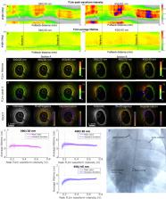
          The clinical management of coronary artery disease and the prevention of acute coronary syndromes require knowledge of the underlying atherosclerotic plaque pathobiology. Hybrid imaging modalities capable of comprehensive assessment of biochemical and morphological plaques features can address this need. Here we report the first implementation of an intravascular catheter system combining fluorescence lifetime imaging (FLIm) with polarization-sensitive optical coherence tomography (PSOCT). This system provides multi-scale assessment of plaque structure and composition via high spatial resolution morphology from OCT, polarimetry-derived tissue microstructure, and biochemical composition from FLIm, without requiring any molecular contrast agent. This result was achieved with a low profile (2.7 Fr) double-clad fiber (DCF) catheter and high speed (100 fps B-scan rate, 40 mm/s pullback speed) console. Use of a DCF and broadband rotary junction required extensive optimization to mitigate the reduction in OCT performance originating from additional reflections and multipath artifacts. This challenge was addressed by the development of a broad-band (UV-visible-IR), high return loss (47 dB) rotary junction. We demonstrate in phantoms, ex vivo swine coronary specimens and in vivo swine heart (percutaneous coronary access) that the FLIm-PSOCT catheter system can simultaneously acquire co-registered FLIm data over four distinct spectral bands (380/20 nm, 400/20 nm, 452/45 nm, 540/45 nm) and PSOCT backscattered intensity, birefringence, and depolarization. The unique ability to collect complementary information from tissue (e.g., morphology, extracellular matrix composition, inflammation) with a device suitable for percutaneous coronary intervention offers new opportunities for cardiovascular research and clinical diagnosis.

          Related collections

          Most cited references48

          • Record: found
          • Abstract: found
          • Article: not found

          Heart Disease and Stroke Statistics—2021 Update: A Report From the American Heart Association

          The American Heart Association, in conjunction with the National Institutes of Health, annually reports the most up-to-date statistics related to heart disease, stroke, and cardiovascular risk factors, including core health behaviors (smoking, physical activity, diet, and weight) and health factors (cholesterol, blood pressure, and glucose control) that contribute to cardiovascular health. The Statistical Update presents the latest data on a range of major clinical heart and circulatory disease conditions (including stroke, congenital heart disease, rhythm disorders, subclinical atherosclerosis, coronary heart disease, heart failure, valvular disease, venous disease, and peripheral artery disease) and the associated outcomes (including quality of care, procedures, and economic costs). The American Heart Association, through its Statistics Committee, continuously monitors and evaluates sources of data on heart disease and stroke in the United States to provide the most current information available in the annual Statistical Update. The 2021 Statistical Update is the product of a full year’s worth of effort by dedicated volunteer clinicians and scientists, committed government professionals, and American Heart Association staff members. This year’s edition includes data on the monitoring and benefits of cardiovascular health in the population, an enhanced focus on social determinants of health, adverse pregnancy outcomes, vascular contributions to brain health, the global burden of cardiovascular disease, and further evidence-based approaches to changing behaviors related to cardiovascular disease. Each of the 27 chapters in the Statistical Update focuses on a different topic related to heart disease and stroke statistics. The Statistical Update represents a critical resource for the lay public, policy makers, media professionals, clinicians, health care administrators, researchers, health advocates, and others seeking the best available data on these factors and conditions.
            Bookmark
            • Record: found
            • Abstract: found
            • Article: not found

            Mechanisms of plaque formation and rupture.

            Atherosclerosis causes clinical disease through luminal narrowing or by precipitating thrombi that obstruct blood flow to the heart (coronary heart disease), brain (ischemic stroke), or lower extremities (peripheral vascular disease). The most common of these manifestations is coronary heart disease, including stable angina pectoris and the acute coronary syndromes. Atherosclerosis is a lipoprotein-driven disease that leads to plaque formation at specific sites of the arterial tree through intimal inflammation, necrosis, fibrosis, and calcification. After decades of indolent progression, such plaques may suddenly cause life-threatening coronary thrombosis presenting as an acute coronary syndrome. Most often, the culprit morphology is plaque rupture with exposure of highly thrombogenic, red cell-rich necrotic core material. The permissive structural requirement for this to occur is an extremely thin fibrous cap, and thus, ruptures occur mainly among lesions defined as thin-cap fibroatheromas. Also common are thrombi forming on lesions without rupture (plaque erosion), most often on pathological intimal thickening or fibroatheromas. However, the mechanisms involved in plaque erosion remain largely unknown, although coronary spasm is suspected. The calcified nodule has been suggested as a rare cause of coronary thrombosis in highly calcified and tortious arteries in older individuals. To characterize the severity and prognosis of plaques, several terms are used. Plaque burden denotes the extent of disease, whereas plaque activity is an ambiguous term, which may refer to one of several processes that characterize progression. Plaque vulnerability describes the short-term risk of precipitating symptomatic thrombosis. In this review, we discuss mechanisms of atherosclerotic plaque initiation and progression; how plaques suddenly precipitate life-threatening thrombi; and the concepts of plaque burden, activity, and vulnerability. © 2014 American Heart Association, Inc.
              Bookmark
              • Record: found
              • Abstract: found
              • Article: not found

              Atherosclerotic plaque progression and vulnerability to rupture: angiogenesis as a source of intraplaque hemorrhage.

              Observational studies of necrotic core progression identify intraplaque hemorrhage as a critical factor in atherosclerotic plaque growth and destabilization. The rapid accumulation of erythrocyte membranes causes an abrupt change in plaque substrate characterized by increased free cholesterol within the lipid core and excessive macrophage infiltration. Neoangiogenesis is associated closely with plaque progression, and microvascular incompetence is a likely source of intraplaque hemorrhage. Intimal neovascularization is predominantly thought to arise from the adventitia, where there are a plethora of pre-existing vasa vasorum. In lesions that have early necrotic cores, the majority of vessels invading from the adventitia occur at specific sites of medial wall disruption. A breech in the medial wall likely facilitates the rapid in-growth of microvessels from the adventitia, and exposure to an atherosclerotic environment stimulates abnormal vascular development characterized by disorganized branching and immature endothelial tubes with "leaky" imperfect linings. This network of immature blood vessels is a viable source of intraplaque hemorrhage providing erythrocyte-derived phospholipids and free cholesterol. The rapid change in plaque substrate caused by the excessive accumulation of erythrocytes may promote the transition from a stable to an unstable lesion. This review discusses the potential role of intraplaque vasa vasorum in lesion instability as it relates to plaque rupture.
                Bookmark

                Author and article information

                Journal
                Biomed Opt Express
                Biomed Opt Express
                BOE
                Biomedical Optics Express
                Optica Publishing Group
                2156-7085
                05 March 2024
                01 April 2024
                : 15
                : 4
                : 2114-2132
                Affiliations
                [1 ]Biomedical Engineering, University of California , Davis, CA 95616, USA
                [2 ]Wellman Center for Photomedicine, Massachusetts General Hospital and Harvard Medical School , Boston, MA 02114, USA
                [3 ]Division of Cardiovascular Medicine, UC Davis Health System, University of California-Davis , Sacramento, CA 95817, USA
                Author notes
                [†]

                These authors contributed equally to this work

                Author information
                https://orcid.org/0000-0003-1222-4071
                https://orcid.org/0000-0002-6715-5176
                https://orcid.org/0000-0003-3819-1271
                https://orcid.org/0000-0003-2369-0748
                Article
                516515
                10.1364/BOE.516515
                11019710
                38633060
                03dd757a-20e7-4c0b-931f-ed5b333724d3
                © 2024 Optica Publishing Group

                https://doi.org/10.1364/OA_License_v2#VOR-OA

                © 2024 Optica Publishing Group under the terms of the Optica Open Access Publishing Agreement

                History
                : 19 December 2023
                : 08 February 2024
                : 08 February 2024
                Funding
                Funded by: National Institutes of Health 10.13039/100000002
                Award ID: P41EB-015903
                Award ID: P41EB-032840
                Award ID: R01HL-157712
                Categories
                Article

                Vision sciences
                Vision sciences

                Comments

                Comment on this article