1
views
0
recommends
+1 Recommend
0 collections
    0
    shares
      • Record: found
      • Abstract: found
      • Article: found
      Is Open Access

      Deep cerebral venous abnormalities in premature babies with GMH-IVH: a single-centre retrospective study

      research-article

      Read this article at

      Bookmark
          There is no author summary for this article yet. Authors can add summaries to their articles on ScienceOpen to make them more accessible to a non-specialist audience.

          Abstract

          Purpose

          Germinal matrix haemorrhage/intraventricular haemorrhage (GMH-IVH) is a multifactorial injury with both anatomic and haemodynamic involvement. Normal variants in preterm deep cerebral venous anatomy associated with GMH-IVH have been previously described using MRI susceptibility weighted imaging (SWI). The aims of this study were to use SWI to compare the deep venous systems of a cohort of preterm neonates with various grades of GMH-IVH to a group of age-matched controls without GMH-IVH and to present novel retrospective SWI imaging findings.

          Methods

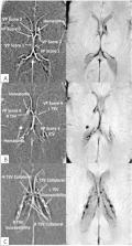
          A neuroradiologist retrospectively evaluated 3T MRI SWI and phase imaging of 56 preterm neonates with GMH-IVH (14 of each grade) and 27 controls without GMH-IVH, scoring the venous irregularities according to three variables: decreased venous patency, increased lumen susceptibility and the presence of collaterals. Eight different venous locations, including indicated bilateral components, were evaluated: straight sinus, vein of galen, internal cerebral, direct lateral, thalamostriate, atrial and the anterior septal veins. Variables were analysed for statistical significance. Inter-rater reliability was determined via subset evaluation by a second paediatric radiologist.

          Results

          Deep venous abnormalities were significantly more common in patients with GMH-IVH, with Wilcoxon Rank Sum Test demonstrating significant increase with GMH-IVH for total decreased venous patency (W=0, p<0.0001), increased lumen susceptibility and collateral formation. Venous abnormalities were also positively correlated with an increase in GMH-IVH grade from I to IV (patency, ρ=0.782, p<0.01) (increased lumen susceptibility, ρ=0.739, p<0.01) (collaterals, ρ=0.649, p<0.01), not just GMH-IVH alone.

          Conclusion

          Deep venous abnormalities are significantly correlated with GMH-IVH alone and an increase in GMH-IVH grade. Further study is needed to determine cause and effect.

          Related collections

          Most cited references38

          • Record: found
          • Abstract: not found
          • Book: not found

          A simple sequentially rejective multiple test procedure

            Bookmark
            • Record: found
            • Abstract: found
            • Article: not found

            Incidence and evolution of subependymal and intraventricular hemorrhage: a study of infants with birth weights less than 1,500 gm.

            We have performed brain scanning by computed tomography on 46 consecutive live-born infants whose birth weights were less than 1,500 gm; 20 of them had evidence of cerebral intraventricular hemorrhage. Nine of the 29 infants who survived had IVH. Four grades of IVH were identified. Grade I and II lesions resolved spontaneously, but there was prominence of the interhemispheric fissue on CT of the infants at six months of age. Hydrocephalus developed in infants with Grade III and IV lesions. Seven of the surviving infants with IVH did not have clinical evidence of hemorrhage. There were no significant differences between the infants with and without IVH in birth weight, gestational age, one- and five-minute Apgar scores, or the need for resuscitation at birth or for subsequent respiratory assistance.
              Bookmark
              • Record: found
              • Abstract: found
              • Article: not found

              The blood-brain barrier: an overview: structure, regulation, and clinical implications.

              The blood-brain barrier (BBB) is a diffusion barrier, which impedes influx of most compounds from blood to brain. Three cellular elements of the brain microvasculature compose the BBB-endothelial cells, astrocyte end-feet, and pericytes (PCs). Tight junctions (TJs), present between the cerebral endothelial cells, form a diffusion barrier, which selectively excludes most blood-borne substances from entering the brain. Astrocytic end-feet tightly ensheath the vessel wall and appear to be critical for the induction and maintenance of the TJ barrier, but astrocytes are not believed to have a barrier function in the mammalian brain. Dysfunction of the BBB, for example, impairment of the TJ seal, complicates a number of neurologic diseases including stroke and neuroinflammatory disorders. We review here the recent developments in our understanding of the BBB and the role of the BBB dysfunction in CNS disease. We have focused on intraventricular hemorrhage (IVH) in premature infants, which may involve dysfunction of the TJ seal as well as immaturity of the BBB in the germinal matrix (GM). A paucity of TJs or PCs, coupled with incomplete coverage of blood vessels by astrocyte end-feet, may account for the fragility of blood vessels in the GM of premature infants. Finally, this review describes the pathogenesis of increased BBB permeability in hypoxia-ischemia and inflammatory mechanisms involving the BBB in septic encephalopathy, HIV-induced dementia, multiple sclerosis, and Alzheimer disease.
                Bookmark

                Author and article information

                Journal
                BMJ Paediatr Open
                BMJ Paediatr Open
                bmjpo
                bmjpo
                BMJ Paediatrics Open
                BMJ Publishing Group (BMA House, Tavistock Square, London, WC1H 9JR )
                2399-9772
                2023
                9 May 2023
                : 7
                : 1
                : e001853
                Affiliations
                [1 ]Ringgold_12233University of Florida College of Medicine , Gainesville, Florida, USA
                [2 ]departmentDepartment of Radiology , Ringgold_12233University of Florida College of Medicine , Gainesville, Florida, USA
                [3 ]departmentDepartment of Mathematics and Statistics , Ringgold_118563Auburn University College of Sciences and Mathematics , Auburn, Alabama, USA
                [4 ]departmentDepartment of Pediatrics , Ringgold_12233University of Florida College of Medicine , Gainesville, Florida, USA
                Author notes
                [Correspondence to ] Dr Thomas Kent; thomaskent@ 123456ufl.edu
                Author information
                http://orcid.org/0000-0001-8834-311X
                Article
                bmjpo-2023-001853
                10.1136/bmjpo-2023-001853
                10174015
                37160379
                03740b64-fa75-4d4d-9a50-66ac888d7bb8
                © Author(s) (or their employer(s)) 2023. Re-use permitted under CC BY-NC. No commercial re-use. See rights and permissions. Published by BMJ.

                This is an open access article distributed in accordance with the Creative Commons Attribution Non Commercial (CC BY-NC 4.0) license, which permits others to distribute, remix, adapt, build upon this work non-commercially, and license their derivative works on different terms, provided the original work is properly cited, appropriate credit is given, any changes made indicated, and the use is non-commercial. See:  http://creativecommons.org/licenses/by-nc/4.0/.

                History
                : 09 January 2023
                : 04 April 2023
                Funding
                Funded by: FundRef http://dx.doi.org/10.13039/100000050, National Heart, Lung, and Blood Institute;
                Award ID: T35HL007489
                Funded by: FundRef http://dx.doi.org/10.13039/100000002, National Institutes of Health;
                Award ID: R01HD105055-01A1
                Categories
                Neonatology
                1506
                Original research
                Custom metadata
                unlocked

                neonatology
                neonatology

                Comments

                Comment on this article