17
views
0
recommends
+1 Recommend
0 collections
    0
    shares
      • Record: found
      • Abstract: found
      • Article: found
      Is Open Access

      TGF‐β signaling directly regulates transcription and functional expression of the electrogenic sodium bicarbonate cotransporter 1, NBCe1 (SLC4A4), via Smad4 in mouse astrocytes

      research-article

      Read this article at

      Bookmark
          There is no author summary for this article yet. Authors can add summaries to their articles on ScienceOpen to make them more accessible to a non-specialist audience.

          Abstract

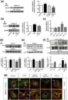
          The electrogenic sodium bicarbonate cotransporter NBCe1 (SLC4A4) expressed in astrocytes regulates intracellular and extracellular pH. Here, we introduce transforming growth factor beta (TGF‐β) as a novel regulator of NBCe1 transcription and functional expression. Using hippocampal slices and primary hippocampal and cortical astrocyte cultures, we investigated regulation of NBCe1 and elucidated the underlying signaling pathways by RT‐PCR, immunoblotting, immunofluorescence, intracellular H( +) recording using the H( +) ‐sensitive dye 2′,7′‐bis‐(carboxyethyl)‐5‐(and‐6)‐carboxyfluorescein, mink lung epithelial cell (MLEC) assay, and chromatin immunoprecipitation. Activation of TGF‐β signaling significantly upregulated transcript, protein, and surface expression of NBCe1. These effects were TGF‐β receptor‐mediated and suppressed following inhibition of JNK and Smad signaling. Moreover, 4‐aminopyridine (4AP)‐dependent NBCe1 regulation requires TGF‐β. TGF‐β increased the rate and amplitude of intracellular H + changes upon challenging NBCe1 in wild‐type astrocytes but not in cortical astrocytes from Slc4a4‐deficient mice. A Smad4 binding sequence was identified in the NBCe1 promoter and Smad4 binding increased after activation of TGF‐β signaling. The data show for the first time that NBCe1 is a direct target of TGF‐β/Smad4 signaling. Through activation of the canonical pathway TGF‐β acts directly on NBCe1 by binding of Smad4 to the NBCe1 promoter and regulating its transcription, followed by increased protein expression and transport activity.

          Related collections

          Most cited references43

          • Record: found
          • Abstract: found
          • Article: not found

          Astrocyte control of synaptic transmission and neurovascular coupling.

          From a structural perspective, the predominant glial cell of the central nervous system, the astrocyte, is positioned to regulate synaptic transmission and neurovascular coupling: the processes of one astrocyte contact tens of thousands of synapses, while other processes of the same cell form endfeet on capillaries and arterioles. The application of subcellular imaging of Ca2+ signaling to astrocytes now provides functional data to support this structural notion. Astrocytes express receptors for many neurotransmitters, and their activation leads to oscillations in internal Ca2+. These oscillations induce the accumulation of arachidonic acid and the release of the chemical transmitters glutamate, d-serine, and ATP. Ca2+ oscillations in astrocytic endfeet can control cerebral microcirculation through the arachidonic acid metabolites prostaglandin E2 and epoxyeicosatrienoic acids that induce arteriole dilation, and 20-HETE that induces arteriole constriction. In addition to actions on the vasculature, the release of chemical transmitters from astrocytes regulates neuronal function. Astrocyte-derived glutamate, which preferentially acts on extrasynaptic receptors, can promote neuronal synchrony, enhance neuronal excitability, and modulate synaptic transmission. Astrocyte-derived d-serine, by acting on the glycine-binding site of the N-methyl-d-aspartate receptor, can modulate synaptic plasticity. Astrocyte-derived ATP, which is hydrolyzed to adenosine in the extracellular space, has inhibitory actions and mediates synaptic cross-talk underlying heterosynaptic depression. Now that we appreciate this range of actions of astrocytic signaling, some of the immediate challenges are to determine how the astrocyte regulates neuronal integration and how both excitatory (glutamate) and inhibitory signals (adenosine) provided by the same glial cell act in concert to regulate neuronal function.
            Bookmark
            • Record: found
            • Abstract: found
            • Article: not found

            Extracellular control of TGFbeta signalling in vascular development and disease.

            The intracellular mechanism of transforming growth factor-beta (TGFbeta) signalling via kinase receptors and SMAD effectors is firmly established, but recent studies of human cardiovascular syndromes such as Marfan syndrome and pre-eclampsia have refocused attention on the importance of regulating the availability of active extracellular TGFbeta. It seems that elastic extracellular matrix (ECM) components have a crucial role in controlling TGFbeta signalling, while soluble and membrane bound forms of TGFbeta co-receptors add further layers of regulation. Together, these extracellular interactions determine the final bioavailability of TGFbeta to vascular cells, and dysregulation is associated with an increasing number of vascular pathologies.
              Bookmark
              • Record: found
              • Abstract: found
              • Article: not found

              An assay for transforming growth factor-beta using cells transfected with a plasminogen activator inhibitor-1 promoter-luciferase construct.

              Transforming growth factor-beta (TGF-beta) is a potent regulator of cellular differentiation, proliferation, migration, and protein expression. These properties have been exploited to create a variety of bioassays for detecting the mature growth factor. In this paper, we describe a highly sensitive and specific, nonradioactive quantitative bioassay for TGF-beta based on its ability to induce plasminogen activator inhibitor-1 (PAI-1) expression. Mink lung epithelial cells (MLEC) were stably transfected with an expression construct containing a truncated PAI-1 promoter fused to the firefly luciferase reporter gene. Addition of TGF-beta (0.2 to > 30 pM) to the transfectants resulted in a dose-dependent increase in luciferase activity in the cell lysates. Although responsive to TGF-beta, this promoter fragment was only minimally influenced by other known inducers of PAI-1 expression. When compared to the widely used MLEC assay, this assay demonstrated greater sensitivity and specificity, allowing quantification of TGF-beta in complex biological solutions.
                Bookmark

                Author and article information

                Contributors
                eleni.roussa@anat.uni-freiburg.de
                Journal
                Glia
                Glia
                10.1002/(ISSN)1098-1136
                GLIA
                Glia
                John Wiley and Sons Inc. (Hoboken )
                0894-1491
                1098-1136
                01 June 2017
                August 2017
                : 65
                : 8 ( doiID: 10.1002/glia.v65.8 )
                : 1361-1375
                Affiliations
                [ 1 ] Department of Molecular Embryology Institute for Anatomy and Cell Biology, Faculty of Medicine, University of Freiburg Albertstrasse 17 D‐79104 Freiburg Germany
                [ 2 ] Department of Neuroanatomy Institute for Anatomy and Cell Biology, Faculty of Medicine, University of Freiburg Albertstrasse 17 D‐79104 Freiburg Germany
                [ 3 ] Department of General Zoology FB Biology, University of Kaiserslautern P.B. 3049 D‐67653 Kaiserslautern Germany
                Author notes
                [*] [* ] Correspondence Dr. Eleni Roussa, Institute for Anatomy and Cell Biology, Department of Molecular Embryology, Faculty of Medicine, University of Freiburg; Albertstrasse 17, D‐79104 Freiburg, Germany. Email: eleni.roussa@ 123456anat.uni-freiburg.de
                Author information
                http://orcid.org/0000-0002-0495-1597
                Article
                GLIA23168
                10.1002/glia.23168
                5518200
                28568893
                019adb72-e08b-4948-bf76-d3c60fde6553
                © 2017 The Authors GLIA Published by Wiley Periodicals, Inc.

                This is an open access article under the terms of the Creative Commons Attribution‐NonCommercial‐NoDerivs License, which permits use and distribution in any medium, provided the original work is properly cited, the use is non‐commercial and no modifications or adaptations are made.

                History
                : 23 January 2017
                : 27 April 2017
                : 04 May 2017
                Page count
                Figures: 6, Tables: 0, Pages: 16, Words: 10095
                Funding
                Funded by: Deutsche Forschungsgemeinschaft
                Award ID: KR1477/15‐1
                Categories
                Research Article
                Research Articles
                Custom metadata
                2.0
                glia23168
                August 2017
                Converter:WILEY_ML3GV2_TO_NLMPMC version:5.1.4 mode:remove_FC converted:20.07.2017

                Neurosciences
                astroglia,epilepsy,growth factor,ph
                Neurosciences
                astroglia, epilepsy, growth factor, ph

                Comments

                Comment on this article