164
views
0
recommends
+1 Recommend
0 collections
    3
    shares
      • Record: found
      • Abstract: found
      • Article: found
      Is Open Access

      Incidence of impacted mandibular and maxillary third molars: a radiographic study in a Southeast Iran population

      research-article

      Read this article at

          There is no author summary for this article yet. Authors can add summaries to their articles on ScienceOpen to make them more accessible to a non-specialist audience.

          Abstract

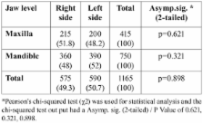
          Objectives: The aim of this study is to evaluate the position of impacted third molars based on the classifications of Pell & Gregory and Winter in a sample of Iranian patients. Study design: In this retrospective study, up to 1020 orthopantomograms (OPG) of the patients who were referred to the radiology clinics from October 2007 to January 2011 were evaluated. Data including the age, gender, the angulation type, width and depth of impaction were evaluated by statistical tests. Results: Among 1020 patients, 380(27.3%) were male and 640(62.7%) were female with the sex ratio was 1:1.7. Of the 1020 OPGs, 585 cases showed at least one impacted third molar, with significant difference between males (205; 35.1%) and females (380; 64.9%) (P = 0.0311). Data analysis showed that impacted third molars were 1.9 times more likely to occur in the mandible than in the maxilla (P =0.000). The most common angulation of impaction in the mandible was mesioangular impaction (48.3%) and the most common angulation of impaction in the maxilla was the vertical (45.3%). Impaction in the level IIA was the most common in both maxilla and mandible. There was no significant diffe-rence between the right and left sides in both the maxilla and the mandible. Conclusion: The pattern of third molar impaction in the southeast region of Iran is characterized by a high prevalence of impaction, especially in the mandible. Female more than male have teeth impaction. The most common angulation was the mesioangular in the mandible, and the vertical angulation in the maxilla. The most common level of impaction was the A and there was no any significant difference between the right and left sides in both jaws.

          Key words:Third molar, impaction, incidence, Iran.

          Related collections

          Most cited references29

          • Record: found
          • Abstract: found
          • Article: not found

          Impacted maxillary canines: a review.

          An overview of the incidence and sequelae, as well as the surgical, periodontal, and orthodontic considerations in the management, of impacted canines is presented. The clinician needs to be familiar with the differences in the surgical management of the labially and palatally impacted canines, the best method of attachment to the canine for orthodontic force application, the advantages of one-arch versus two-arch treatment, and the implications of canine extraction. The various factors that influence all these decisions are discussed.
            Bookmark
            • Record: found
            • Abstract: found
            • Article: not found

            Pattern of third molar impaction in a Singapore Chinese population: a retrospective radiographic survey.

            One thousand orthopantomograms (OPGs) of patients 20-40 years old were examined. Where impacted third molars were present, the angle and depth of impaction were recorded. Results were analysed using the Pearson chi2 test. 68.6% of OPGs showed at least one impacted third molar. The frequency was three-fold higher in the mandible (1024/1079=90%) than in the maxilla (306/1077=28%), with a significantly higher frequency (P<0.05) in females (56%) than males (44%). The mesioangular impaction was the most common, and 80% of all impacted third molars were partially buried in bone. Of the 429 bilateral occurrence of impacted third molars, 423 were in the mandible. It was concluded that the frequency of impacted third molars in the Singapore Chinese population studied was generally two to three times that reported in races of the Caucasian stock. There was also double the frequency of impacted third molars when compared to a previous study in a Chinese population published in 1932 with females being more frequently affected than males.
              Bookmark
              • Record: found
              • Abstract: found
              • Article: not found

              Third molar outcomes from age 18 to 26: findings from a population-based New Zealand longitudinal study.

              The purpose of this study was to describe the presence and impaction status of people's third molars at age 18 years, as well as the observed changes in their clinical status between ages 18 and 26 years. Eight hundred twenty-one participants in a prospective cohort study were dentally examined at ages 18 and 26 years, and panoramic radiographs were taken at age 18 years but not at 26 years. For each tooth, its radiographic impaction status at age 18 years was compared with the clinical status by age 26 years. Of the 2857 third molars assessed at age 18 years, 92.8% were able to be followed up clinically at age 26 years. Approximately 54.9% of the teeth that were not impacted by age 18 had erupted by 26 years. Of the teeth that were impacted by age 18, 33.7% had fully erupted by age 26, 31.4% had been extracted, and 13.1% remained unerupted. Of the maxillary teeth that were categorized as "impacted" at age 18 years, 36.2% had fully erupted by age 26, whereas only 25.6% of the mandibular teeth had done so (P <.01). Fewer mandibular teeth than maxillary teeth remained unerupted by the time the patient was 26 years old (27.4% and 41.4%, respectively; P <.01), but there was no significant difference between the jaws in the proportion of impacted teeth at age 18 years that had been extracted by age 26 years (29.6% and 30.3%, respectively). For mesioangularly impacted third molars, 39.3% of maxillary teeth and 20.4% of mandibular teeth had fully erupted by age 26. Of the third molars that were mesioangularly impacted at age 18 years, 39.3% of maxillary teeth and 20.4% of mandibular teeth had fully erupted by age 26 years, whereas almost one-third of each had been extracted. Of the distoangularly impacted third molars, 20.4% of the maxillary teeth and one-third of the mandibular teeth had erupted by age 26, with 21.6% of the maxillary teeth and 31.6% of the mandibular teeth having been extracted. Other than horizontally impacted third molars, a substantial proportion of other impaction types do erupt fully, and radiographically apparent impaction in late adolescence should not be sufficient grounds for their prophylactic removal in the absence of other clinical indications.
                Bookmark

                Author and article information

                Journal
                Med Oral Patol Oral Cir Bucal
                Med Oral Patol Oral Cir Bucal
                Medicina Oral S.L.
                Medicina Oral, Patología Oral y Cirugía Bucal
                Medicina Oral S.L.
                1698-4447
                1698-6946
                January 2013
                10 December 2012
                : 18
                : 1
                : e140-e145
                Affiliations
                [1 ]DDS, MSc. Member of Kerman Dental and Oral Diseases Research Center. Department of Oral Medicine, School of Dentistry, Kerman University of Medical Sciences, Kerman, Iran
                [2 ]DDS. Member of Kerman dental and oral diseases research center. Kerman University of Medical Sciences, Kerman, Iran
                [3 ]DDS, MSc. Department of Radiology, University of Medical Sciences, Kerman, Iran
                Author notes
                Department of Oral Medicine School of Dentistry Kerman University of Medical Sciences Kerman, Iran , E-mail: m_hashemipoor@ 123456kmu.ac.ir
                Article
                18028
                10.4317/medoral.18028
                3548634
                23229243
                63d708a6-f568-44d5-861e-1dff36a56c4b
                Copyright: © 2013 Medicina Oral S.L.

                This is an open-access article distributed under the terms of the Creative Commons Attribution License, which permits unrestricted use, distribution, and reproduction in any medium, provided the original work is properly cited.

                History
                : 7 June 2012
                : 23 October 2011
                Categories
                Research-Article
                Clinical and Experimental dentistry

                Surgery
                Surgery

                Comments

                Comment on this article