11
views
0
recommends
+1 Recommend
0 collections
    0
    shares
      • Record: found
      • Abstract: found
      • Article: not found

      Activatable molecular probes for fluorescence-guided surgery, endoscopy and tissue biopsy

      Read this article at

      ScienceOpenPublisher
          There is no author summary for this article yet. Authors can add summaries to their articles on ScienceOpen to make them more accessible to a non-specialist audience.

          Abstract

          We highlight the development of activatable molecular probes that trigger the optical signals toward biomarkers, allowing real-time, dynamic visualization of lesions and margins for guided-surgery, endoscopy and tissue biopsy with molecular precision.

          Abstract

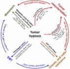
          The real-time, dynamic optical visualization of lesions and margins ensures not only complete resection of the malignant tissues but also better preservation of the vital organs/tissues during surgical procedures. Most imaging probes with an “always-on” signal encounter high background noise due to their non-specific accumulation in normal tissues. By contrast, activatable molecular probes only “turn on” their signals upon reaction with the targeted biomolecules that are overexpressed in malignant cells, offering high target-to-background ratios with high specificity and sensitivity. This review summarizes the recent progress of activatable molecular probes in surgical imaging and diagnosis. The design principle and mechanism of activatable molecular probes are discussed, followed by specific emphasis on applications ranging from fluorescence-guided surgery to endoscopy and tissue biopsy. Finally, potential challenges and perspectives in the field of activatable molecular probe-enabled surgical imaging are discussed.

          Related collections

          Most cited references174

          • Record: found
          • Abstract: found
          • Article: not found

          Artificial intelligence in radiology

          Artificial intelligence (AI) algorithms, particularly deep learning, have demonstrated remarkable progress in image-recognition tasks. Methods ranging from convolutional neural networks to variational autoencoders have found myriad applications in the medical image analysis field, propelling it forward at a rapid pace. Historically, in radiology practice, trained physicians visually assessed medical images for the detection, characterization and monitoring of diseases. AI methods excel at automatically recognizing complex patterns in imaging data and providing quantitative, rather than qualitative, assessments of radiographic characteristics. In this O pinion article, we establish a general understanding of AI methods, particularly those pertaining to image-based tasks. We explore how these methods could impact multiple facets of radiology, with a general focus on applications in oncology, and demonstrate ways in which these methods are advancing the field. Finally, we discuss the challenges facing clinical implementation and provide our perspective on how the domain could be advanced.
            Bookmark
            • Record: found
            • Abstract: found
            • Article: not found

            Aggregation-induced emission.

            Luminogenic materials with aggregation-induced emission (AIE) attributes have attracted much interest since the debut of the AIE concept in 2001. In this critical review, recent progress in the area of AIE research is summarized. Typical examples of AIE systems are discussed, from which their structure-property relationships are derived. Through mechanistic decipherment of the photophysical processes, structural design strategies for generating new AIE luminogens are developed. Technological, especially optoelectronic and biological, applications of the AIE systems are exemplified to illustrate how the novel AIE effect can be utilized for high-tech innovations (183 references). This journal is © The Royal Society of Chemistry 2011
              Bookmark
              • Record: found
              • Abstract: found
              • Article: found
              Is Open Access

              The role of hypoxia in cancer progression, angiogenesis, metastasis, and resistance to therapy

              Hypoxia is a non-physiological level of oxygen tension, a phenomenon common in a majority of malignant tumors. Tumor-hypoxia leads to advanced but dysfunctional vascularization and acquisition of epithelial-to-mesenchymal transition phenotype resulting in cell mobility and metastasis. Hypoxia alters cancer cell metabolism and contributes to therapy resistance by inducing cell quiescence. Hypoxia stimulates a complex cell signaling network in cancer cells, including the HIF, PI3K, MAPK, and NFĸB pathways, which interact with each other causing positive and negative feedback loops and enhancing or diminishing hypoxic effects. This review provides background knowledge on the role of tumor hypoxia and the role of the HIF cell signaling involved in tumor blood vessel formation, metastasis, and development of the resistance to therapy. Better understanding of the role of hypoxia in cancer progression will open new windows for the discovery of new therapeutics targeting hypoxic tumor cells and hypoxic microenvironment.
                Bookmark

                Author and article information

                Contributors
                Journal
                CSRVBR
                Chemical Society Reviews
                Chem. Soc. Rev.
                Royal Society of Chemistry (RSC)
                0306-0012
                1460-4744
                January 24 2022
                2022
                : 51
                : 2
                : 566-593
                Affiliations
                [1 ]National Engineering Research Centre for Nanomedicine, College of Life Science and Technology, Huazhong University of Science and Technology, Wuhan 430074, China
                [2 ]Hubei Key Laboratory of Bioinorganic Chemistry and Materia Medical, Huazhong University of Science and Technology, Wuhan 430074, China
                [3 ]Department of Nuclear Medicine, Tongji Hospital, Tongji Medical College, Huazhong University of Science and Technology, Wuhan 430030, P. R. China
                [4 ]School of Chemical and Biomedical Engineering, Nanyang Technological University, 70 Nanyang Drive, 637457, Singapore
                Article
                10.1039/D1CS00525A
                f0a7d88f-af16-4462-a57a-79b382fcde72
                © 2022

                http://rsc.li/journals-terms-of-use

                History

                Comments

                Comment on this article