3
views
0
recommends
+1 Recommend
0 collections
    0
    shares
      • Record: found
      • Abstract: found
      • Article: found
      Is Open Access

      Imaging of pituitary tumors: an update with the 5th WHO Classifications—part 2. Neoplasms other than PitNET and tumor-mimicking lesions

      review-article
      ,
      Japanese Journal of Radiology
      Springer Nature Singapore
      Pituitary tumors, WHO classification, Diagnostic imaging, MRI

      Read this article at

          There is no author summary for this article yet. Authors can add summaries to their articles on ScienceOpen to make them more accessible to a non-specialist audience.

          Abstract

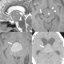
          Many types of tumors can develop in the pituitary gland. In the recently revised 5th editions of the World Health Organization (WHO) classifications (2021 WHO Classification of Central Nervous System Tumors and the 2022 WHO Classification of Endocrine and Neuroendocrine Tumors), various changes have been made to the tumors other than pituitary neuroendocrine tumor (PitNET)/pituitary adenoma, as well as PitNET. Adamantinomatous craniopharyngioma and papillary craniopharyngioma are now considered separate tumors in the 5th edition of the WHO classification. Tumors positive for thyroid transcription factor 1, a marker of posterior pituitary cells, are now grouped together in the pituicyte tumor family in the 5th edition of the WHO classification of Endocrine and Neuroendocrine Tumors. Poorly differentiated chordoma is newly listed in the 5th edition of the WHO Classification of Endocrine and Neuroendocrine Tumors. In this paper, we present the latest WHO classification of pituitary tumors (adamantinomatous craniopharyngioma, papillary craniopharyngioma, pituitary blastoma, pituicyte tumor family, tumors of pituitary origin other than those of the pituicyte tumor family, germinoma, meningioma, chordoma, metastatic tumors, lymphoma, and pituitary incidentaloma), review diseases requiring differentiation from tumors (pituitary abscess, hypophysitis, pituitary hyperplasia, Rathke’s cleft cyst, arachnoid cyst, and aneurysm), and discuss diagnoses based on imaging findings.

          Related collections

          Most cited references107

          • Record: found
          • Abstract: found
          • Article: not found

          Incidence of Endocrine Dysfunction Following the Use of Different Immune Checkpoint Inhibitor Regimens

          If not promptly recognized, endocrine dysfunction can be life threatening. The incidence and risk of developing such adverse events (AEs) following the use of immune checkpoint inhibitor (ICI) regimens are unknown.
            Bookmark
            • Record: found
            • Abstract: found
            • Article: not found

            Chordoma: incidence and survival patterns in the United States, 1973-1995.

            Chordoma, a rare tumor arising from notochordal remnants, has been described to date only by single-institution case series or small population-based surveys. We used data from the Surveillance, Epidemiology, and End Results (SEER) program of the National Cancer Institute, 1973-1995, to calculate age-adjusted incidence and survival rates for 400 cases of microscopically confirmed chordoma and to derive information regarding case distribution and risk of second cancer. The age-adjusted chordoma incidence rate (IR) of 0.08 per 100,000 was age-dependent, more common in males (IR 0.10) than females (IR 0.06) and rare among patients aged <40 years and blacks. Within the axial skeleton 32% of cases were cranial, 32.8% spinal and 29.2% sacral. Young age (<26 years; p = 0.0001) and female sex (p = 0.037) were associated with greater likelihood of cranial presentation. There was no overall increased risk for second primary cancers after chordoma. Median survival was 6.29 years; 5- and 10-year relative survival rates were 67.6% and 39.9%, respectively. Comparison with other bone sarcomas revealed racial disparities in incidence for the two developmental tumors, chordoma and Ewing's sarcoma. This study provides new data regarding incidence and survival patterns of chordoma in the US. Additional epidemiologic studies are required to elucidate the genetic and environmental determinants underlying this rare, distinctive neoplasm.
              Bookmark
              • Record: found
              • Abstract: found
              • Article: not found

              Autoimmune hypophysitis.

              Autoimmune (lymphocytic) hypophysitis is a rare disease that should be considered in the differential diagnosis of any nonsecreting pituitary mass, especially when occurring during pregnancy or postpartum. We have analyzed 370 articles published from January 1962 to October 2004 and identified a total of 379 patients with primary lymphocytic hypophysitis. The present review synthesizes the clinical and research data reported in this body of scientific literature.
                Bookmark

                Author and article information

                Contributors
                yukiomiki0615-ca@yahoo.co.jp
                Journal
                Jpn J Radiol
                Jpn J Radiol
                Japanese Journal of Radiology
                Springer Nature Singapore (Singapore )
                1867-1071
                1867-108X
                13 March 2023
                13 March 2023
                2023
                : 41
                : 8
                : 808-829
                Affiliations
                GRID grid.518217.8, ISNI 0000 0005 0893 4200, Department of Diagnostic and Interventional Radiology, Graduate School of Medicine, , Osaka Metropolitan University, ; 1-4-3 Asahi-Machi, Abeno-Ku, Osaka, 545-8585 Japan
                Author information
                http://orcid.org/0000-0003-0621-0044
                Article
                1407
                10.1007/s11604-023-01407-0
                10366287
                36913010
                c9ad1d82-a4a1-4e6a-9280-430fa5f38c60
                © The Author(s) 2023, corrected publication 2023

                Open AccessThis article is licensed under a Creative Commons Attribution 4.0 International License, which permits use, sharing, adaptation, distribution and reproduction in any medium or format, as long as you give appropriate credit to the original author(s) and the source, provide a link to the Creative Commons licence, and indicate if changes were made. The images or other third party material in this article are included in the article's Creative Commons licence, unless indicated otherwise in a credit line to the material. If material is not included in the article's Creative Commons licence and your intended use is not permitted by statutory regulation or exceeds the permitted use, you will need to obtain permission directly from the copyright holder. To view a copy of this licence, visit http://creativecommons.org/licenses/by/4.0/.

                History
                : 25 December 2022
                : 24 February 2023
                Categories
                Invited Review
                Custom metadata
                © Japan Radiological Society 2023

                pituitary tumors,who classification,diagnostic imaging,mri

                Comments

                Comment on this article