6
views
0
recommends
+1 Recommend
3 collections
    0
    shares
      • Record: found
      • Abstract: found
      • Article: found

      Evans syndrome and immune thrombocytopenia in two patients with COVID‐19

      letter

      Read this article at

      ScienceOpenPublisherPMC
          There is no author summary for this article yet. Authors can add summaries to their articles on ScienceOpen to make them more accessible to a non-specialist audience.

          Abstract

          Dear Editor, 1 1.1 The COVID‐19 pandemic caused by the SARS‐CoV‐2 virus has enveloped the globe with 83 million cases and 1,831,703 deaths worldwide, at the time of writing. 1 Among the hematological manifestations described, severe and symptomatic thrombocytopenia has been rare. A meta‐analysis of 7613 patients found platelet counts to be much lower in patients with severe COVID‐19. 2 Here, we report on two patients with COVID‐19, one with Evans syndrome and one with immune thrombocytopenia to highlight the rarer hematological manifestations of the disease. As there is an association between thrombocytopenia and higher mortality, early identification and treatment could potentially improve outcomes. Patient 1: A 33‐year‐old man presented to the emergency department with a 3‐week history of gum bleeding, black tarry stools, and reddish spots on the skin. He had no fever, cough, or dyspnea. On examination, he had petechial lesions over the chest, legs, and oral mucosa. Laboratory investigations revealed severe thrombocytopenia with initial platelet counts of 6 × 109/L. The peripheral smear showed 11 nucleated RBCs per 100 WBCs, poikilocytosis, ovalocytes, and polychromatic cells with no schistocytes. He had leucocytosis (12 × 109/L), anemia (7.5 g/dl), and elevated lactate dehydrogenase 1953 U/L (normal range, 225–460 U/L). Total and direct bilirubin were 1.23 and 0.46, mean corpuscular volume was 86.8 pl, mean corpuscular hemoglobin 28.3 pg, mean corpuscular hemoglobin concentration 32.6%, and reticulocyte count was 13.73% (corrected 6.87%). Direct Coombs test was positive (2+), suggesting immune hemolytic anemia. Within a few hours of admission, the patient complained of sudden‐onset headache and developed a generalized tonic–clonic seizure. Computed tomography of the brain showed intracerebral hemorrhage in the right capsuloganglionic region with edema and midline shift. The patient's sensorium worsened rapidly with anisocoria, and he was shifted to the intensive care unit. Serology for dengue and scrub typhus (common regional causes of thrombocytopenia) were negative. Nasopharyngeal swab reverse‐transcription polymerase chain reaction (RT‐PCR) for SARS‐CoV‐2 was positive. Immune destruction being the likely cause, he was treated with pulse dexamethasone 40 mg daily with platelet transfusions (intravenous immunoglobulin [IVIG] was not feasible). Bone marrow aspiration was not done. Despite the above measures, there was no improvement in the patient's platelet counts (Figure 1) nor sensorium, and he died on the third day of admission. He had not received anticoagulation. Figure 1 Thrombocytopenia trend during admission Patient 2: A 54‐year‐old man presented to the emergency department with low‐grade intermittent fever and sore throat for a week. He tested positive for COVID‐19 by nasopharyngeal swab RT‐PCR. His initial platelet count was 80 × 109/L, with hemoglobin 15 g/dl and a total leukocyte count of 2.8 × 109/L. Dengue and scrub typhus serology were negative. During the admission, his platelet count dropped to 30 × 109/L. He did not have any bleeding manifestations. He was treated with dexamethasone 6 mg daily from admission, for COVID‐19, based on existing hospital protocol at the time. Platelet counts recovered to 105 × 109/L over a week (Figure 1). His symptoms subsided by Day 3, and he was discharged on Day 8. He received no anticoagulation. These cases suggest an association between COVID‐19, Evans syndrome, and immune thrombocytopenia, based on temporal profile and other etiologies having been ruled out to a reasonable extent. They also highlight heterogeneity in the hematological manifestations of COVID‐19 ranging from asymptomatic thrombocytopenia to life‐threatening disease. This is the second case of Evans syndrome with COVID‐19 described in the literature to the best of our knowledge. Li et al. 3 have reported immune thrombocytopenia and hemolytic anemia in a 39‐year‐old man with COVID‐19. Following IVIG treatment, he improved. Zulfiqar et al. 4 have described a patient with COVID‐19 and immune thrombocytopenia, who developed a subarachnoid hemorrhage. The patient was initiated on IVIG at admission, and the hemorrhage occurred on Day 9. Bomhof et al. 5 described a patient like ours with immune thrombocytopenia associated with COVID‐19 who died following intracerebral hemorrhage. The largest series of seven patients with autoimmune hemolytic anemia, without thrombocytopenia, has been described by Lazarian et al. 6 Nearly all patients were treated with steroids, and two required rituximab. 
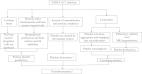
The mechanisms suggested for thrombocytopenia include SARS‐CoV‐2 entering hematopoietic cells via the CD13 receptors causing aberrant hematopoiesis, 7 immune destruction due to molecular mimicry between platelet membrane components (especially glycoprotein) and virus antigens, 8 and increased consumption due to endothelial injury and microangiopathy. The commonest mechanism described in the literature so far has been immune‐mediated destruction (Table 1), which was likely the cause in both of our patients as well. It is imperative to anticipate this complication for early diagnosis and initiation of therapy, as mortality is high. Table 1 Case reports and series of patients with COVID‐19 and severe thrombocytopenia Author Number of patients Month Country Nadir platelet count Clinical bleeding Treatment Outcome Possible mechanism Zulfiqar et al. 4 1 April 2020 France 8 × 109/L Petechiae, subarachnoid hemorrhage Prednisolone (high dose) Alive Immune destruction Deruelle et al. 9 1 May 2020 France 19 × 109/L Tracheal bleed Intravenous methylprednisolone Alive Immune destruction Bomhof et al. 5 3 July 2020 Netherlands 2 × 109/L P1‐skin, mucosal petechiae; P2‐ petechiae, epistaxis; P3‐ intracerebral bleed IVIG, dexamethasone P1‐alive; P2‐alive; P3‐died Immune destruction Humbert et al. 8 1 August 2020 France 4 × 109/L Hematuria, epistaxis Prednisolone, IVIG Not mentioned Immune destruction Ahmed et al. 10 3 June 2020 United Kingdom 0 × 109/L P1‐epistaxis, petechiae; P2‐ petechiae, epistaxis; P3‐ intracerebral bleed IVIG P1‐alive; P2‐alive; P3‐died Immune destruction Lingamaneni et al. 11 1 Jan 2020 USA 96 × 109/L Nil Heparin withheld Died Heparin‐induced thrombocytopenia Li et al. 3 1 June 2020 USA 3 × 109/L Hematemesis, melena, hematochezia IVIG Alive Immune destruction (Evans syndrome) Mahevas et al. 12 14 Aug 2020 France <1 × 109/L Purpura, mucosal, GI bleeding, epistaxis IVIG/steroids Alive Immune thrombocytopenia Current study 2 July 2020 India 4 × 109/L 30 × 109/L P1‐intracerebral bleed P2‐no bleed Dexamethasone Died Alive P1‐immune destruction (Evans syndrome) P2‐immune destruction Abbreviation: IVIG, intravenous immunoglobulin. John Wiley & Sons, Ltd. This article is being made freely available through PubMed Central as part of the COVID-19 public health emergency response. It can be used for unrestricted research re-use and analysis in any form or by any means with acknowledgement of the original source, for the duration of the public health emergency. CONFLICT OF INTERESTS The authors declare that there are no conflict of interests. AUTHOR CONTRIBUTIONS Josh T. Georgy: Conceptualization; methodology; formal analysis; investigation; writing—original draft; visualization. Jonathan A. J. Jayakaran: Conceptualization; methodology; formal analysis; investigation; writing—review, and editing; visualization. Anju S. Jacob: Conceptualization; methodology; formal analysis; investigation; writing—review, and editing; visualization. Karthik Gunasekaran: Conceptualization; methodology; formal analysis; investigation; writing—review and editing, visualization. Pritish J. Korula: Methodology; formal analysis; writing—review, and editing; visualization. Anup J. Devasia: Formal analysis; investigation; writing—review, and editing; visualization. Ramya Iyadurai: Writing—review, and editing; visualization; supervision; project administration; resources.

          Related collections

          Most cited references11

          • Record: found
          • Abstract: found
          • Article: found
          Is Open Access

          Mechanism of thrombocytopenia in COVID-19 patients

          Since December 2019, a novel coronavirus has spread throughout China and across the world, causing a continuous increase in confirmed cases within a short period of time. Some studies reported cases of thrombocytopenia, but hardly any studies mentioned how the virus causes thrombocytopenia. We propose several mechanisms by which coronavirus disease 2019 causes thrombocytopenia to better understand this disease and provide more clinical treatment options.
            Bookmark
            • Record: found
            • Abstract: found
            • Article: not found

            Immune Thrombocytopenic Purpura in a Patient with Covid-19

            To rapidly communicate information on the global clinical effort against Covid-19, the Journal has initiated a series of case reports that offer important teaching points or novel findings. The case reports should be viewed as observations rather than as recommendations for evaluation or treatment. In the interest of timeliness, these reports are evaluated by in-house editors, with peer review reserved for key points as needed. A 65-year-old woman with hypertension, autoimmune hypothyroidism, and known Covid-19 exposure presented to the emergency department with a 4-day history of fatigue, fever, dry cough, and abdominal discomfort. She was afebrile and had a respiratory rate of 28 breaths per minute and an oxygen saturation of 89% while she was breathing ambient air; breath sounds were diminished bilaterally with bibasilar rales. The abdominal examination was normal. Laboratory tests showed a normal white-cell count, hemoglobin level (14.2 g per deciliter), and platelet count (183,000 per cubic millimeter). The C-reactive protein level was elevated at 55 mg per liter, and liver-function tests showed cholestasis. An oropharyngeal swab for Covid-19 testing was positive. Chest computed tomography (CT) showed ground-glass opacities in the lower zones. The patient was admitted and began to receive treatment with intravenous amoxicillin–clavulanic acid, low-molecular-weight heparin, and oxygen. On day 4, lower-extremity purpura (Figure 1) appeared and epistaxis occurred. Isolated thrombocytopenia (platelet count, 66,000 per cubic millimeter) was noted; these findings prompted discontinuation of heparin and antibiotics. On day 5, the platelet count was 16,000 per cubic millimeter, and on day 7, it was 8000 per cubic millimeter. The prothrombin and activated partial thromboplastin times were normal; the fibrinogen level was 5 g per liter (normal range, 2 to 4). A peripheral-blood smear showed less than 1% schistocytes. The level of thyroid peroxidase antibodies was 245 U per milliliter (normal value, <35); antiplatelet factor 4, antiplatelet antibodies, and antinuclear antibodies were not detected. Intravenous immune globulin was administered at a rate of 1 g per kilogram of body weight. After 48 hours, the platelet count decreased to 1000 per cubic millimeter and the purpuric lesions progressed; this prompted a second infusion of immune globulin at a dose of 1 g per kilogram. 1,2 Bone marrow aspiration showed normal cellularity with an increase in pleomorphic megakaryocytes. Erythroblast and granulocyte cell lines were normal, without evidence of hemophagocytosis. On day 9, a right frontal headache developed in the patient, without fever, vomiting, or focal neurologic deficit. CT of the head showed a subarachnoid microhemorrhage in the right frontal lobe. The platelet count was 2000 per cubic millimeter, and a platelet transfusion was administered with 100 mg of prednisolone; eltrombopag (75 mg per day) was initiated. 1,2 On day 10, the headache had resolved, there were no new neurologic findings, and the platelet count had increased to 10,000 per cubic millimeter. All the other laboratory tests had normalized except for the fibrinogen level, which remained elevated (3.5 g per liter). On day 13, the platelet count was 139,000 per cubic millimeter, and the purpura had disappeared. The temporal sequence in this case suggests, 3 but does not prove, that Covid-19 was a causal factor in immune thrombocytopenia in this patient. Fortunately, she had a response to treatment, albeit in a delayed fashion. Although the cerebral bleeding did not have major sequelae, this case illustrates the need to be vigilant for complications associated with Covid-19.
              Bookmark
              • Record: found
              • Abstract: found
              • Article: found

              Autoimmune haemolytic anaemia associated with COVID‐19 infection

              Among patients with SARS‐CoV‐2 infection (also known as COVID‐19), pneumonia, respiratory failure and acute respiratory distress syndrome are frequently encountered complications. 1 Although the pathophysiology underlying severe COVID‐19 remains poorly understood, accumulating evidence argues for hyperinflammatory syndrome causing fulminant and fatal cytokines release associated with disease severity and poor outcome. 2 However, the spectrum of complications is broader and includes among others various auto‐immune disorders such as autoimmune thrombocytopenia, Guillain–Barré and antiphospholipid syndrome. 3 , 4 , 5 In this report we describe seven patients from six French and Belgian Hospitals who developed a first episode of autoimmune haemolytic anaemia (AIHA) during a COVID‐19 infection. Patient characteristics are detailed in Table I. Briefly, median age was 62 years (range, 61–89 years), and all patients presented with risk factors for developing a severe form of COVID‐19 such as hypertension, diabetes and chronic renal failure. All patients had both a positive oropharyngeal swab for SARS‐CoV‐2 and typical images of COVID‐19 infection on chest computed tomography scans (25–75% extension). Three patients were admitted in an intensive care unit but only one required invasive ventilation. Treatment for COVID‐19 infection differed according to the standards of each centre. Thus, three patients received hydroxychloroquine, in association with azithromycin for two of them, and one patient received lopinavir and ritonavir. Table I Characteristics of seven patients with autoimmune haemolytic anaemia after the onset of COVID‐19. Patient Age Gender Comorbidity CT‐scan* Oropharyngeal swab (tested by PCR) Haemoglobin (g/l) Reticulocyte count (109/l) Lymphocyte count (109/l) Lactate dehydrogenase (U/l) Haptoglobin (g/l) DAT specificity Optimum temperature Day between COVID‐19 symptoms and AIHA Related pathology AIHA treatment Response #1 61 M Hypertension, chronic renal failure Moderate Positive 60 477 250 1000 100 g/l along with an increase of 20 g/l at least seven days after an infusion with red blood cells. Corticosteroid failure lead to rituximab injection in the third case (patient #6), and one responding patient is scheduled to receive rituximab because of a MZL clone (patient #3). At the time of last follow‐up, all patients were alive and had at least partly recovered from COVID‐19. To conclude, we report seven cases of warm and cold AIHA associated with COVID‐19 disease, all of them occurring after the beginning of the symptoms of the infection and within a timeframe compatible with that of the cytokine storm. Four out of the seven patients had indolent B lymphoid malignancy either already known or discovered because of the haemolytic episode. AIHA is a classical complication of both CLL and MZL, 6 , 7 and viral infections are known to trigger autoimmune cytopenias. 8 Whether the presence of an underlying malignant B lymphoid clone facilitated the onset of AIHA is unknown. Nonetheless, these observations argue for systematically investigating for the presence of a lymphoid clone in patients presenting with COVID‐19 infections and autoimmune cytopenias. Author contributions GL, AQ and FC designed the research study, analyzed the data and wrote the paper. MB, JS, CJ, DR, FM, AM, TB, GD and AD contributed to conception, patient enrollment and data collection.
                Bookmark

                Author and article information

                Contributors
                joshtg@gmail.com , jtgeorgy@cmcvellore.ac.in , med5@cmcvellore.ac.in
                Journal
                J Med Virol
                J Med Virol
                10.1002/(ISSN)1096-9071
                JMV
                Journal of Medical Virology
                John Wiley and Sons Inc. (Hoboken )
                0146-6615
                1096-9071
                09 March 2021
                : 10.1002/jmv.26906
                Affiliations
                [ 1 ] Department of Medicine Unit V Christian Medical College Vellore Tamil Nadu India
                [ 2 ] Department of Critical Care Christian Medical College Vellore Tamil Nadu India
                [ 3 ] Department of Haematology Christian Medical College Vellore Tamil Nadu India
                Author notes
                [*] [* ] Correspondence Josh T. Georgy, Department of Medicine Unit V, Christian Medical College and Hospital, Vellore, Tamil Nadu 632004, India.

                Email: joshtg@ 123456gmail.com , jtgeorgy@ 123456cmcvellore.ac.in and med5@ 123456cmcvellore.ac.in

                Author information
                http://orcid.org/0000-0002-7119-9710
                Article
                JMV26906
                10.1002/jmv.26906
                8013600
                33634495
                e88f490f-6410-41ac-b7a4-85721bde555f
                © 2021 Wiley Periodicals LLC

                This article is being made freely available through PubMed Central as part of the COVID-19 public health emergency response. It can be used for unrestricted research re-use and analysis in any form or by any means with acknowledgement of the original source, for the duration of the public health emergency.

                History
                : 29 January 2021
                : 08 December 2020
                : 24 February 2021
                Page count
                Figures: 1, Tables: 1, Pages: 3, Words: 1381
                Categories
                Letter to the Editor
                Letter to the Editor
                Custom metadata
                2.0
                corrected-proof
                Converter:WILEY_ML3GV2_TO_JATSPMC version:6.0.1 mode:remove_FC converted:01.04.2021

                Microbiology & Virology
                Microbiology & Virology

                Comments

                Comment on this article