6
views
0
recommends
+1 Recommend
0 collections
    0
    shares
      • Record: found
      • Abstract: found
      • Article: found
      Is Open Access

      Identification of Small Nucleolar RNA SNORD60 as a Potential Biomarker and Its Clinical Significance in Lung Adenocarcinoma

      research-article

      Read this article at

          There is no author summary for this article yet. Authors can add summaries to their articles on ScienceOpen to make them more accessible to a non-specialist audience.

          Abstract

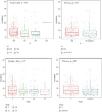
          Non-small-cell lung cancer (NSCLC) is the leading cause of cancer deaths in the world and often diagnosed at an advanced stage, so it is urgent to explore the pathogenesis and new diagnostic biomarkers. Accumulated evidences suggested that small nucleolar RNAs (snoRNAs) played a key role in the development and progression of NSCLC. To examine differential expression snoRNA profile and identify snoRNAs with clinical significance in lung adenocarcinoma (LUAD), The Cancer Genome Atlas (TCGA) LUAD RNA sequencing dataset was used to investigate differential expression snoRNA signatures and compared with snoRNA PCR array analysis in pair-matched LUAD tissues. The diagnostic ability of SONRD60 was assessed using a receiver operating characteristic (ROC) curve. The Kaplan-Meier method was used to plot survival curves. Univariate and multivariate Cox regression analyses were used to investigate the prognostic effect of SNORD60 expression on LUAD. The results showed that SNORD60 was a significantly upregulated snoRNA after intersection analysis in LUAD cases. SNORD60 has 74.2% sensitivity and 75.3% specificity for the diagnosis of LUAD. Increased SNORD60 expression was linked with lymph node metastases and the TNM stage ( P < 0.05). Pathological T category and lymph node metastases were independent prognostic factors for overall survival in a multivariate Cox regression study. Our findings demonstrated that SNORD60, a small nucleolar RNA, has an oncogenic function in LUAD and might be used as a new early diagnostic biomarker for LUAD.

          Related collections

          Most cited references36

          • Record: found
          • Abstract: found
          • Article: not found

          Global cancer statistics 2020: GLOBOCAN estimates of incidence and mortality worldwide for 36 cancers in 185 countries

          This article provides an update on the global cancer burden using the GLOBOCAN 2020 estimates of cancer incidence and mortality produced by the International Agency for Research on Cancer. Worldwide, an estimated 19.3 million new cancer cases (18.1 million excluding nonmelanoma skin cancer) and almost 10.0 million cancer deaths (9.9 million excluding nonmelanoma skin cancer) occurred in 2020. Female breast cancer has surpassed lung cancer as the most commonly diagnosed cancer, with an estimated 2.3 million new cases (11.7%), followed by lung (11.4%), colorectal (10.0 %), prostate (7.3%), and stomach (5.6%) cancers. Lung cancer remained the leading cause of cancer death, with an estimated 1.8 million deaths (18%), followed by colorectal (9.4%), liver (8.3%), stomach (7.7%), and female breast (6.9%) cancers. Overall incidence was from 2-fold to 3-fold higher in transitioned versus transitioning countries for both sexes, whereas mortality varied <2-fold for men and little for women. Death rates for female breast and cervical cancers, however, were considerably higher in transitioning versus transitioned countries (15.0 vs 12.8 per 100,000 and 12.4 vs 5.2 per 100,000, respectively). The global cancer burden is expected to be 28.4 million cases in 2040, a 47% rise from 2020, with a larger increase in transitioning (64% to 95%) versus transitioned (32% to 56%) countries due to demographic changes, although this may be further exacerbated by increasing risk factors associated with globalization and a growing economy. Efforts to build a sustainable infrastructure for the dissemination of cancer prevention measures and provision of cancer care in transitioning countries is critical for global cancer control.
            Bookmark
            • Record: found
            • Abstract: found
            • Article: not found

            The 2015 World Health Organization Classification of Lung Tumors: Impact of Genetic, Clinical and Radiologic Advances Since the 2004 Classification.

            The 2015 World Health Organization (WHO) Classification of Tumors of the Lung, Pleura, Thymus and Heart has just been published with numerous important changes from the 2004 WHO classification. The most significant changes in this edition involve (1) use of immunohistochemistry throughout the classification, (2) a new emphasis on genetic studies, in particular, integration of molecular testing to help personalize treatment strategies for advanced lung cancer patients, (3) a new classification for small biopsies and cytology similar to that proposed in the 2011 Association for the Study of Lung Cancer/American Thoracic Society/European Respiratory Society classification, (4) a completely different approach to lung adenocarcinoma as proposed by the 2011 Association for the Study of Lung Cancer/American Thoracic Society/European Respiratory Society classification, (5) restricting the diagnosis of large cell carcinoma only to resected tumors that lack any clear morphologic or immunohistochemical differentiation with reclassification of the remaining former large cell carcinoma subtypes into different categories, (6) reclassifying squamous cell carcinomas into keratinizing, nonkeratinizing, and basaloid subtypes with the nonkeratinizing tumors requiring immunohistochemistry proof of squamous differentiation, (7) grouping of neuroendocrine tumors together in one category, (8) adding NUT carcinoma, (9) changing the term sclerosing hemangioma to sclerosing pneumocytoma, (10) changing the name hamartoma to "pulmonary hamartoma," (11) creating a group of PEComatous tumors that include (a) lymphangioleiomyomatosis, (b) PEComa, benign (with clear cell tumor as a variant) and
              Bookmark
              • Record: found
              • Abstract: found
              • Article: found
              Is Open Access

              Comprehensive molecular profiling of lung adenocarcinoma

              Adenocarcinoma of the lung is the leading cause of cancer death worldwide. Here we report molecular profiling of 230 resected lung adenocarcinomas using messenger RNA, microRNA and DNA sequencing integrated with copy number, methylation and proteomic analyses. High rates of somatic mutation were seen (mean 8.9 mutations per megabase). Eighteen genes were statistically significantly mutated, including RIT1 activating mutations and newly described loss-of-function MGA mutations which are mutually exclusive with focal MYC amplification. EGFR mutations were more frequent in female patients, whereas mutations in RBM10 were more common in males. Aberrations in NF1, MET, ERBB2 and RIT1 occurred in 13% of cases and were enriched in samples otherwise lacking an activated oncogene, suggesting a driver role for these events in certain tumours. DNA and mRNA sequence from the same tumour highlighted splicing alterations driven by somatic genomic changes, including exon 14 skipping in MET mRNA in 4% of cases. MAPK and PI(3)K pathway activity, when measured at the protein level, was explained by known mutations in only a fraction of cases, suggesting additional, unexplained mechanisms of pathway activation. These data establish a foundation for classification and further investigations of lung adenocarcinoma molecular pathogenesis.
                Bookmark

                Author and article information

                Contributors
                Journal
                Biomed Res Int
                Biomed Res Int
                BMRI
                BioMed Research International
                Hindawi
                2314-6133
                2314-6141
                2022
                7 June 2022
                : 2022
                : 5501171
                Affiliations
                1Chinese PLA Medical School, Beijing 100853, China
                2Department of General Medicine, The Fourth Medical Center of PLA General Hospital, Beijing 100048, China
                3Department of Oncology, Air Force Medical Center, PLA, Beijing 100142, China
                4Hebei North University, Zhangjiakou 075000, Hebei Province, China
                5Department of Thoracic Surgery, The Fourth Medical Center of PLA General Hospital, Beijing 100048, China
                6Senior Department of Oncology, The Fifth Medical Center of PLA General Hospital, Beijing 100071, China
                7Department of Thoracic Surgery, Beijing Aerospace General Hospital, Beijing 100076, China
                Author notes

                Academic Editor: Qiang Liu

                Author information
                https://orcid.org/0000-0002-1388-5608
                https://orcid.org/0000-0002-4146-5918
                https://orcid.org/0000-0003-2594-5871
                https://orcid.org/0000-0002-0426-6290
                https://orcid.org/0000-0001-8767-7898
                https://orcid.org/0000-0002-7025-735X
                https://orcid.org/0000-0003-3523-2760
                https://orcid.org/0000-0001-8986-6027
                Article
                10.1155/2022/5501171
                9197630
                a8fca8ef-8474-4177-931c-3dda270bfa4c
                Copyright © 2022 Hongwei Zhou et al.

                This is an open access article distributed under the Creative Commons Attribution License, which permits unrestricted use, distribution, and reproduction in any medium, provided the original work is properly cited.

                History
                : 7 April 2022
                : 6 May 2022
                Funding
                Funded by: Air Force Medical Center
                Award ID: 21ZT15
                Funded by: National Natural Science Foundation of China
                Award ID: 81601984
                Categories
                Research Article

                Comments

                Comment on this article