23
views
0
recommends
+1 Recommend
0 collections
    0
    shares
      • Record: found
      • Abstract: found
      • Article: found
      Is Open Access

      Astrocytes convert network excitation to tonic inhibition of neurons

      research-article

      Read this article at

          There is no author summary for this article yet. Authors can add summaries to their articles on ScienceOpen to make them more accessible to a non-specialist audience.

          Abstract

          Background

          Glutamate and γ-aminobutyric acid (GABA) transporters play important roles in balancing excitatory and inhibitory signals in the brain. Increasing evidence suggest that they may act concertedly to regulate extracellular levels of the neurotransmitters.

          Results

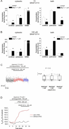
          Here we present evidence that glutamate uptake-induced release of GABA from astrocytes has a direct impact on the excitability of pyramidal neurons in the hippocampus. We demonstrate that GABA, synthesized from the polyamine putrescine, is released from astrocytes by the reverse action of glial GABA transporter (GAT) subtypes GAT-2 or GAT-3. GABA release can be prevented by blocking glutamate uptake with the non-transportable inhibitor DHK, confirming that it is the glutamate transporter activity that triggers the reversal of GABA transporters, conceivably by elevating the intracellular Na + concentration in astrocytes. The released GABA significantly contributes to the tonic inhibition of neurons in a network activity-dependent manner. Blockade of the Glu/GABA exchange mechanism increases the duration of seizure-like events in the low-[Mg 2+] in vitro model of epilepsy. Under in vivo conditions the increased GABA release modulates the power of gamma range oscillation in the CA1 region, suggesting that the Glu/GABA exchange mechanism is also functioning in the intact hippocampus under physiological conditions.

          Conclusions

          The results suggest the existence of a novel molecular mechanism by which astrocytes transform glutamat ergic excitation into GABA ergic inhibition providing an adjustable, in situ negative feedback on the excitability of neurons.

          Related collections

          Most cited references74

          • Record: found
          • Abstract: found
          • Article: not found

          Glutamate uptake.

          Brain tissue has a remarkable ability to accumulate glutamate. This ability is due to glutamate transporter proteins present in the plasma membranes of both glial cells and neurons. The transporter proteins represent the only (significant) mechanism for removal of glutamate from the extracellular fluid and their importance for the long-term maintenance of low and non-toxic concentrations of glutamate is now well documented. In addition to this simple, but essential glutamate removal role, the glutamate transporters appear to have more sophisticated functions in the modulation of neurotransmission. They may modify the time course of synaptic events, the extent and pattern of activation and desensitization of receptors outside the synaptic cleft and at neighboring synapses (intersynaptic cross-talk). Further, the glutamate transporters provide glutamate for synthesis of e.g. GABA, glutathione and protein, and for energy production. They also play roles in peripheral organs and tissues (e.g. bone, heart, intestine, kidneys, pancreas and placenta). Glutamate uptake appears to be modulated on virtually all possible levels, i.e. DNA transcription, mRNA splicing and degradation, protein synthesis and targeting, and actual amino acid transport activity and associated ion channel activities. A variety of soluble compounds (e.g. glutamate, cytokines and growth factors) influence glutamate transporter expression and activities. Neither the normal functioning of glutamatergic synapses nor the pathogenesis of major neurological diseases (e.g. cerebral ischemia, hypoglycemia, amyotrophic lateral sclerosis, Alzheimer's disease, traumatic brain injury, epilepsy and schizophrenia) as well as non-neurological diseases (e.g. osteoporosis) can be properly understood unless more is learned about these transporter proteins. Like glutamate itself, glutamate transporters are somehow involved in almost all aspects of normal and abnormal brain activity.
            Bookmark
            • Record: found
            • Abstract: found
            • Article: not found

            Astrocyte control of synaptic transmission and neurovascular coupling.

            From a structural perspective, the predominant glial cell of the central nervous system, the astrocyte, is positioned to regulate synaptic transmission and neurovascular coupling: the processes of one astrocyte contact tens of thousands of synapses, while other processes of the same cell form endfeet on capillaries and arterioles. The application of subcellular imaging of Ca2+ signaling to astrocytes now provides functional data to support this structural notion. Astrocytes express receptors for many neurotransmitters, and their activation leads to oscillations in internal Ca2+. These oscillations induce the accumulation of arachidonic acid and the release of the chemical transmitters glutamate, d-serine, and ATP. Ca2+ oscillations in astrocytic endfeet can control cerebral microcirculation through the arachidonic acid metabolites prostaglandin E2 and epoxyeicosatrienoic acids that induce arteriole dilation, and 20-HETE that induces arteriole constriction. In addition to actions on the vasculature, the release of chemical transmitters from astrocytes regulates neuronal function. Astrocyte-derived glutamate, which preferentially acts on extrasynaptic receptors, can promote neuronal synchrony, enhance neuronal excitability, and modulate synaptic transmission. Astrocyte-derived d-serine, by acting on the glycine-binding site of the N-methyl-d-aspartate receptor, can modulate synaptic plasticity. Astrocyte-derived ATP, which is hydrolyzed to adenosine in the extracellular space, has inhibitory actions and mediates synaptic cross-talk underlying heterosynaptic depression. Now that we appreciate this range of actions of astrocytic signaling, some of the immediate challenges are to determine how the astrocyte regulates neuronal integration and how both excitatory (glutamate) and inhibitory signals (adenosine) provided by the same glial cell act in concert to regulate neuronal function.
              Bookmark
              • Record: found
              • Abstract: found
              • Article: not found

              Sulforhodamine 101 as a specific marker of astroglia in the neocortex in vivo.

              Glial cells have been identified as key signaling components in the brain; however, methods to investigate their structure and function in vivo have been lacking. Here, we describe a new, highly selective approach for labeling astrocytes in intact rodent neocortex that allows in vivo imaging using two-photon microscopy. The red fluorescent dye sulforhodamine 101 (SR101) was specifically taken up by protoplasmic astrocytes after brief exposure to the brain surface. Specificity was confirmed by immunohistochemistry. In addition, SR101 labeled enhanced green fluorescent protein (EGFP)-expressing astrocytes but not microglial cells in transgenic mice. We used SR101 labeling to quantify morphological characteristics of astrocytes and to visualize their close association with the cortical microvasculature. Furthermore, by combining this method with calcium indicator loading of cell populations, we demonstrated distinct calcium dynamics in astroglial and neuronal networks. We expect SR101 staining to become a principal tool for investigating astroglia in vivo.
                Bookmark

                Author and article information

                Journal
                BMC Biol
                BMC Biol
                BMC Biology
                BioMed Central
                1741-7007
                2012
                15 March 2012
                : 10
                : 26
                Affiliations
                [1 ]Department of Functional Pharmacology, Institute of Molecular Pharmacology, Research Centre for Natural Sciences, Hungarian Academy of Sciences, Pusztaszeri 59-67, 1025 Budapest, Hungary
                [2 ]Laboratory of Neuromorphology and Neuroendocrinology, Semmelweis University and Hungarian Academy of Sciences, Tűzoltó 58, 1094 Budapest, Hungary
                [3 ]Department of Biochemical Pharmacology, Institute of Molecular Pharmacology, Research Centre for Natural Sciences, Hungarian Academy of Sciences, Pusztaszeri 59-67, 1025 Budapest, Hungary
                [4 ]Comparative Psychophysiology Group, Institute of Cognitive Neuroscience and Psychology, Research Centre for Natural Sciences, Hungarian Academy of Sciences, Victor Hugo 18-22, 1132 Budapest, Hungary
                [5 ]Péter Pázmány Catholic University, Faculty of Information Technology, Práter 50A, 1083 Budapest, Hungary
                Article
                1741-7007-10-26
                10.1186/1741-7007-10-26
                3342137
                22420899
                9d3e4c76-6350-43c5-bdc7-6959eaf03c32
                Copyright ©2012 Héja et al; licensee BioMed Central Ltd.

                This is an Open Access article distributed under the terms of the Creative Commons Attribution License ( http://creativecommons.org/licenses/by/2.0), which permits unrestricted use, distribution, and reproduction in any medium, provided the original work is properly cited.

                History
                : 20 December 2011
                : 15 March 2012
                Categories
                Research Article

                Life sciences
                Life sciences

                Comments

                Comment on this article