15
views
0
recommends
+1 Recommend
0 collections
    0
    shares
      • Record: found
      • Abstract: found
      • Article: found
      Is Open Access

      Quantification of severe liver iron overload using MRI offset echoes

      case-report

      Read this article at

          There is no author summary for this article yet. Authors can add summaries to their articles on ScienceOpen to make them more accessible to a non-specialist audience.

          Abstract

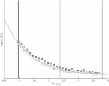
          Magnetic resonance imaging (MRI) has become the clinical standard to estimate liver iron overload. The most commonly used method is to measure the transversal relaxation time, T2*, from a multi gradient recalled echo sequence (MGRE). While this technique is reliable in low to moderate liver iron concentrations (LIC), it will be inaccurate when it is severe. We report a case with severe liver hemochromatosis and show the benefit of using an easily implemented MRI offset echo sequence to more accurately estimate LIC. After adjusting treatment, both Ferritin and LIC decreased. Using standard MGRE this reduction could not have been detected.

          Related collections

          Most cited references6

          • Record: found
          • Abstract: found
          • Article: found
          Is Open Access

          Biopsy-based calibration of T2* magnetic resonance for estimation of liver iron concentration and comparison with R2 Ferriscan

          Background There is a need to standardise non-invasive measurements of liver iron concentrations (LIC) so clear inferences can be drawn about body iron levels that are associated with hepatic and extra-hepatic complications of iron overload. Since the first demonstration of an inverse relationship between biopsy LIC and liver magnetic resonance (MR) using a proof-of-concept T2* sequence, MR technology has advanced dramatically with a shorter minimum echo-time, closer inter-echo spacing and constant repetition time. These important advances allow more accurate calculation of liver T2* especially in patients with high LIC. Methods Here, we used an optimised liver T2* sequence calibrated against 50 liver biopsy samples on 25 patients with transfusional haemosiderosis using ordinary least squares linear regression, and assessed the method reproducibility in 96 scans over an LIC range up to 42 mg/g dry weight (dw) using Bland-Altman plots. Using mixed model linear regression we compared the new T2*-LIC with R2-LIC (Ferriscan) on 92 scans in 54 patients with transfusional haemosiderosis and examined method agreement using Bland-Altman approach. Results Strong linear correlation between ln(T2*) and ln(LIC) led to the calibration equation LIC = 31.94(T2*)-1.014. This yielded LIC values approximately 2.2 times higher than the proof-of-concept T2* method. Comparing this new T2*-LIC with the R2-LIC (Ferriscan) technique in 92 scans, we observed a close relationship between the two methods for values up to 10 mg/g dw, however the method agreement was poor. Conclusions New calibration of T2* against liver biopsy estimates LIC in a reproducible way, correcting the proof-of-concept calibration by 2.2 times. Due to poor agreement, both methods should be used separately to diagnose or rule out liver iron overload in patients with increased ferritin.
            Bookmark
            • Record: found
            • Abstract: found
            • Article: not found

            Magnetic resonance imaging quantification of liver iron.

            Iron overload is the histologic hallmark of hereditary hemochromatosis and transfusional hemosiderosis but also may occur in chronic hepatopathies. This article provides an overview of iron deposition and diseases where liver iron overload is clinically relevant. Next, this article reviews why quantitative noninvasive biomarkers of liver iron would be beneficial. Finally, we describe current state-of-the-art methods for quantifying iron with MR imaging and review remaining challenges and unsolved problems. Copyright © 2010 Elsevier Inc. All rights reserved.
              Bookmark
              • Record: found
              • Abstract: found
              • Article: not found

              Effect of multipeak spectral modeling of fat for liver iron and fat quantification: correlation of biopsy with MR imaging results.

              To investigate the effect of the multipeak spectral modeling of fat on R2* values as measures of liver iron and on the quantification of liver fat fraction, with biopsy as the reference standard. Institutional review board approval and informed consent were obtained. Patients with liver disease (n = 95; 50 men, 45 women; mean age, 57.2 years±14.1 [standard deviation]) underwent a nontargeted liver biopsy, and 97 biopsy samples were reviewed for steatosis and iron grades. MR imaging at 1.5 T was performed 24-72 hours after biopsy by using a three-echo three-dimensional gradient-echo sequence for water and fat separation. Data were reconstructed off-line, correcting for T1 and T2* effects. Fat fraction and R2* maps (1/T2*) were reconstructed and differences in R2* and steatosis grades with and without multipeak modeling of fat were tested by using the Kruskal-Wallis test. Spearman rank correlation coefficient was used to assess fat fractions and steatosis grades. Linear regression analysis was performed to compare the fat fraction for both models. Mean steatosis grade at biopsy ranged from 0% to 95%. Biopsy specimens in 26 of 97 patients (27%) showed liver iron (15 mild, six moderate, and five severe). In all 71 samples without iron, a strong increase in the apparent R2* was observed with increasing steatosis grade when single-peak modeling of fat was used (P=.001). When multipeak modeling was used, there were no differences in the apparent R2* as a function of steatosis grading (P=.645), and R2* values agreed closely with those reported in the literature. Good correlation between fat fraction and steatosis grade was observed (rS=0.85) both without and with spectral modeling. In the presence of fat, multipeak spectral modeling of fat improves the agreement between R2* and liver iron. Single-peak modeling of fat leads to underestimation of liver fat. © RSNA, 2012.
                Bookmark

                Author and article information

                Journal
                Acta Radiol Open
                Acta Radiol Open
                ARR
                sparr
                Acta Radiologica Open
                SAGE Publications (Sage UK: London, England )
                2058-4601
                25 May 2015
                May 2015
                : 4
                : 5
                : 2047981614568910
                Affiliations
                [1 ]Department of Medical Physics, Karolinska University Hospital, Stockholm, Sweden
                [2 ]Department of Molecular Medicine and Surgery, Karolinska Institute, Stockholm, Sweden
                [3 ]Department of Radiology, Karolinska University Hospital, Stockholm, Sweden
                Author notes
                [*]Mikael Skorpil, Department of Radiology, Karolinska University Hospital, Solna 17176, Sweden. Email: mikael.skorpil@ 123456ki.se
                Article
                10.1177_2047981614568910
                10.1177/2047981614568910
                4446225
                36846013-1460-43d4-8457-127dab8093e1
                © The Foundation Acta Radiologica 2015 Reprints and permissions: sagepub.co.uk/journalsPermissions.nav

                This article is distributed under the terms of the Creative Commons Attribution-NonCommercial 3.0 License ( http://www.creativecommons.org/licenses/by-nc/3.0/) which permits non-commercial use, reproduction and distribution of the work without further permission provided the original work is attributed as specified on the SAGE and Open Access page( http://www.uk.sagepub.com/aboutus/openaccess.htm).

                History
                : 27 August 2014
                : 30 December 2014
                Categories
                Case Report
                Custom metadata
                corrected-proof

                abdomen/gi,magnetic resonance imaging (mri),liver,adults and pediatrics,imaging sequences

                Comments

                Comment on this article