28
views
0
recommends
+1 Recommend
0 collections
    0
    shares
      • Record: found
      • Abstract: found
      • Article: found
      Is Open Access

      Coupling of Polo kinase activation to nuclear localization by a bifunctional NLS is required during mitotic entry

      research-article

      Read this article at

          There is no author summary for this article yet. Authors can add summaries to their articles on ScienceOpen to make them more accessible to a non-specialist audience.

          Abstract

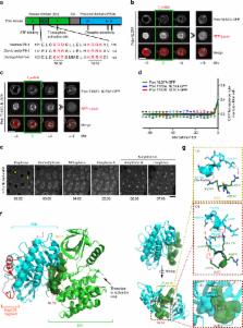
          The Polo kinase is a master regulator of mitosis and cytokinesis conserved from yeasts to humans. Polo is composed of an N-term kinase domain (KD) and a C-term polo-box domain (PBD), which regulates its subcellular localizations. The PBD and KD can interact and inhibit each other, and this reciprocal inhibition is relieved when Polo is phosphorylated at its activation loop. How Polo activation and localization are coupled during mitotic entry is unknown. Here we report that PBD binding to the KD masks a nuclear localization signal (NLS). Activating phosphorylation of the KD leads to exposure of the NLS and entry of Polo into the nucleus before nuclear envelope breakdown. Failures of this mechanism result in misregulation of the Cdk1-activating Cdc25 phosphatase and lead to mitotic and developmental defects in Drosophila. These results uncover spatiotemporal mechanisms linking master regulatory enzymes during mitotic entry.

          Abstract

          Drosophila Polo kinase is the founding member of the Polo-Like Kinase (PLK) family and a master regulator of mitosis and cytokinesis. Here the authors uncover a molecular mechanism for the spatiotemporal regulation of Polo kinase during mitotic entry through a phosphorylation event that triggers nuclear import.

          Related collections

          Most cited references44

          • Record: found
          • Abstract: found
          • Article: not found

          Protein kinases: evolution of dynamic regulatory proteins.

          Eukayotic protein kinases evolved as a family of highly dynamic molecules with strictly organized internal architecture. A single hydrophobic F-helix serves as a central scaffold for assembly of the entire molecule. Two non-consecutive hydrophobic structures termed "spines" anchor all the elements important for catalysis to the F-helix. They make firm, but flexible, connections within the molecule, providing a high level of internal dynamics of the protein kinase. During the course of evolution, protein kinases developed a universal regulatory mechanism associated with a large activation segment that can be dynamically folded and unfolded in the course of cell functioning. Protein kinases thus represent a unique, highly dynamic, and precisely regulated set of switches that control most biological events in eukaryotic cells. Copyright © 2010. Published by Elsevier Ltd.
            Bookmark
            • Record: found
            • Abstract: found
            • Article: not found

            Regulation of protein kinases; controlling activity through activation segment conformation.

            There are currently at least forty-six unique protein kinase crystal structures, twenty-four of which are available in an active state. Here we examine these structures using a structural bioinformatics approach to understand how the conformation of the activation segment controls kinase activity. Copyright 2004 Cell Press
              Bookmark
              • Record: found
              • Abstract: found
              • Article: not found

              The molecular basis for phosphodependent substrate targeting and regulation of Plks by the Polo-box domain.

              Polo-like kinases (Plks) perform crucial functions in cell-cycle progression and multiple stages of mitosis. Plks are characterized by a C-terminal noncatalytic region containing two tandem Polo boxes, termed the Polo-box domain (PBD), which has recently been implicated in phosphodependent substrate targeting. We show that the PBDs of human, Xenopus, and yeast Plks all recognize similar phosphoserine/threonine-containing motifs. The 1.9 A X-ray structure of a human Plk1 PBD-phosphopeptide complex shows that the Polo boxes each comprise beta6alpha structures that associate to form a 12-stranded beta sandwich domain. The phosphopeptide binds along a conserved, positively charged cleft located at the edge of the Polo-box interface. Mutations that specifically disrupt phosphodependent interactions abolish cell-cycle-dependent localization and provide compelling phenotypic evidence that PBD-phospholigand binding is necessary for proper mitotic progression. In addition, phosphopeptide binding to the PBD stimulates kinase activity in full-length Plk1, suggesting a conformational switching mechanism for Plk regulation and a dual functionality for the PBD.
                Bookmark

                Author and article information

                Contributors
                vincent.archambault.1@umontreal.ca
                Journal
                Nat Commun
                Nat Commun
                Nature Communications
                Nature Publishing Group UK (London )
                2041-1723
                22 November 2017
                22 November 2017
                2017
                : 8
                : 1701
                Affiliations
                [1 ]ISNI 0000 0001 2292 3357, GRID grid.14848.31, Institute for Research in Immunology and Cancer, Université de Montréal, C.P. 6128 Succursale Centre-Ville, ; Montréal, QC Canada H3C 3J7
                [2 ]ISNI 0000 0001 2292 3357, GRID grid.14848.31, Département de biochimie et médecine moléculaire, , Université de Montréal, C.P. 6128 Succursale Centre-Ville, ; Montréal, QC Canada H3C 3J7
                Article
                1876
                10.1038/s41467-017-01876-8
                5700101
                fc369a7d-d73c-4e34-a624-ccf1577aeb88
                © The Author(s) 2017

                Open Access This article is licensed under a Creative Commons Attribution 4.0 International License, which permits use, sharing, adaptation, distribution and reproduction in any medium or format, as long as you give appropriate credit to the original author(s) and the source, provide a link to the Creative Commons license, and indicate if changes were made. The images or other third party material in this article are included in the article’s Creative Commons license, unless indicated otherwise in a credit line to the material. If material is not included in the article’s Creative Commons license and your intended use is not permitted by statutory regulation or exceeds the permitted use, you will need to obtain permission directly from the copyright holder. To view a copy of this license, visit http://creativecommons.org/licenses/by/4.0/.

                History
                : 5 May 2017
                : 22 October 2017
                Categories
                Article
                Custom metadata
                © The Author(s) 2017

                Uncategorized
                Uncategorized

                Comments

                Comment on this article