13
views
0
recommends
+1 Recommend
0 collections
    0
    shares
      • Record: found
      • Abstract: found
      • Article: found
      Is Open Access

      Phase separation by the polyhomeotic sterile alpha motif compartmentalizes Polycomb Group proteins and enhances their activity

      research-article

      Read this article at

      Bookmark
          There is no author summary for this article yet. Authors can add summaries to their articles on ScienceOpen to make them more accessible to a non-specialist audience.

          Abstract

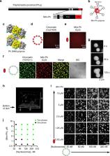
          Polycomb Group (PcG) proteins organize chromatin at multiple scales to regulate gene expression. A conserved Sterile Alpha Motif (SAM) in the Polycomb Repressive Complex 1 (PRC1) subunit Polyhomeotic (Ph) has been shown to play an important role in chromatin compaction and large-scale chromatin organization. Ph SAM forms helical head to tail polymers, and SAM-SAM interactions between chromatin-bound Ph/PRC1 are believed to compact chromatin and mediate long-range interactions. To understand the underlying mechanism, here we analyze the effects of Ph SAM on chromatin in vitro. We find that incubation of chromatin or DNA with a truncated Ph protein containing the SAM results in formation of concentrated, phase-separated condensates. Ph SAM-dependent condensates can recruit PRC1 from extracts and enhance PRC1 ubiquitin ligase activity towards histone H2A. We show that overexpression of Ph with an intact SAM increases ubiquitylated H2A in cells. Thus, SAM-induced phase separation, in the context of Ph, can mediate large-scale compaction of chromatin into biochemical compartments that facilitate histone modification.

          Abstract

          The conserved SAM motif of Polycomb Repressive Complex 1 subunit Ph has been shown to play an important role in chromatin organization. Here, the authors study the effect of Ph SAM on chromatin in vitro, showing that it induces the formation of concentrated, phase-separated condensates, which enhance the ubiquitin ligase activity of PRC1.

          Related collections

          Most cited references86

          • Record: found
          • Abstract: found
          • Article: not found

          Biomolecular condensates: organizers of cellular biochemistry

          In addition to membrane-bound organelles, eukaryotic cells feature various membraneless compartments, including the centrosome, the nucleolus and various granules. Many of these compartments form through liquid–liquid phase separation, and the principles, mechanisms and regulation of their assembly as well as their cellular functions are now beginning to emerge.
            Bookmark
            • Record: found
            • Abstract: found
            • Article: not found

            Protein Phase Separation: A New Phase in Cell Biology

            Cellular compartments and organelles organize biological matter. Most well-known organelles are separated by a membrane boundary from their surrounding milieu. There are also many so-called membraneless organelles and recent studies suggest that these organelles, which are supramolecular assemblies of proteins and RNA molecules, form via protein phase separation. Recent discoveries have shed light on the molecular properties, formation, regulation, and function of membraneless organelles. A combination of techniques from cell biology, biophysics, physical chemistry, structural biology, and bioinformatics are starting to help establish the molecular principles of an emerging field, thus paving the way for exciting discoveries, including novel therapeutic approaches for the treatment of age-related disorders.
              Bookmark
              • Record: found
              • Abstract: found
              • Article: not found

              Transcription Factors Activate Genes through the Phase-Separation Capacity of Their Activation Domains

              Gene expression is controlled by transcription factors (TFs) that consist of DNA-binding domains (DBDs) and activation domains (ADs). The DBDs have been well-characterized, but little is known about the mechanisms by which ADs effect gene activation. Here we report that diverse ADs form phase-separated condensates with the Mediator coactivator. For the OCT4 and GCN4 TFs, we show that the ability to form phase-separated droplets with Mediator in vitro and the ability to activate genes in vivo are dependent on the same amino acid residues. For the estrogen receptor (ER), a ligand-dependent activator, we show that estrogen enhances phase separation with Mediator, again linking phase separation with gene activation. These results suggest that diverse TFs can interact with Mediator through the phase-separating capacity of their ADs and that formation of condensates with Mediator is involved in gene activation. Activation domains from a diverse array of mammalian and yeast transcription factors form phase-separated condensates with mediator to activate gene expression
                Bookmark

                Author and article information

                Contributors
                nicole.francis@ircm.qc.ca
                Journal
                Nat Commun
                Nat Commun
                Nature Communications
                Nature Publishing Group UK (London )
                2041-1723
                5 November 2020
                5 November 2020
                2020
                : 11
                : 5609
                Affiliations
                [1 ]GRID grid.14848.31, ISNI 0000 0001 2292 3357, Institut de recherches cliniques de Montréal, ; 110 Avenue des Pins Ouest, Montréal, QC H2W 1R7 Canada
                [2 ]GRID grid.14709.3b, ISNI 0000 0004 1936 8649, Division of Experimental Medicine, , McGill University, ; 1001 Decarie Boulevard, Montreal, QC H4A 3J1 Canada
                [3 ]GRID grid.260024.2, Department of Biochemistry and Molecular Genetics, , Midwestern University, ; 19555N. 59th St., Glendale, AZ 85308 USA
                [4 ]GRID grid.14848.31, ISNI 0000 0001 2292 3357, Département de biochimie et médecine moléculaire Université de Montréal, ; 2900 Boulevard Edouard-Montpetit, Montréal, QC H3T 1J4 Canada
                Author information
                http://orcid.org/0000-0002-1810-2959
                Article
                19435
                10.1038/s41467-020-19435-z
                7644731
                33154383
                d4224bcc-d0b5-4df7-bd8e-56d3a1187c2d
                © The Author(s) 2020

                Open Access This article is licensed under a Creative Commons Attribution 4.0 International License, which permits use, sharing, adaptation, distribution and reproduction in any medium or format, as long as you give appropriate credit to the original author(s) and the source, provide a link to the Creative Commons license, and indicate if changes were made. The images or other third party material in this article are included in the article’s Creative Commons license, unless indicated otherwise in a credit line to the material. If material is not included in the article’s Creative Commons license and your intended use is not permitted by statutory regulation or exceeds the permitted use, you will need to obtain permission directly from the copyright holder. To view a copy of this license, visit http://creativecommons.org/licenses/by/4.0/.

                History
                : 25 February 2020
                : 8 October 2020
                Funding
                Funded by: FundRef https://doi.org/10.13039/100000057, U.S. Department of Health & Human Services | NIH | National Institute of General Medical Sciences (NIGMS);
                Award ID: GM114338-02
                Award Recipient :
                Categories
                Article
                Custom metadata
                © The Author(s) 2020

                Uncategorized
                enzyme mechanisms,ligases,ubiquitylated proteins,ubiquitylation,histone post-translational modifications

                Comments

                Comment on this article