13
views
0
recommends
+1 Recommend
0 collections
    0
    shares
      • Record: found
      • Abstract: found
      • Article: found
      Is Open Access

      To be and not to be: wide-field Ca 2+ imaging reveals neocortical functional segmentation combines stability and flexibility

      research-article

      Read this article at

          There is no author summary for this article yet. Authors can add summaries to their articles on ScienceOpen to make them more accessible to a non-specialist audience.

          Abstract

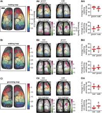
          The stability and flexibility of the functional parcellation of the cerebral cortex is fundamental to how familiar and novel information is both represented and stored. We leveraged new advances in Ca 2+ sensors and microscopy to understand the dynamics of functional segmentation in the dorsal cerebral cortex. We performed wide-field Ca 2+ imaging in head-fixed mice and used spatial independent component analysis (ICA) to identify independent spatial sources of Ca 2+ fluorescence. The imaging data were evaluated over multiple timescales and discrete behaviors including resting, walking, and grooming. When evaluated over the entire dataset, a set of template independent components (ICs) were identified that were common across behaviors. Template ICs were present across a range of timescales, from days to 30 seconds, although with lower occurrence probability at shorter timescales, highlighting the stability of the functional segmentation. Importantly, unique ICs emerged at the shorter duration timescales that could act to transiently refine the cortical network. When data were evaluated by behavior, both common and behavior-specific ICs emerged. Each behavior is composed of unique combinations of common and behavior-specific ICs. These observations suggest that cerebral cortical functional segmentation exhibits considerable spatial stability over time and behaviors while retaining the flexibility for task-dependent reorganization.

          Related collections

          Most cited references88

          • Record: found
          • Abstract: found
          • Article: not found

          The human brain is intrinsically organized into dynamic, anticorrelated functional networks.

          During performance of attention-demanding cognitive tasks, certain regions of the brain routinely increase activity, whereas others routinely decrease activity. In this study, we investigate the extent to which this task-related dichotomy is represented intrinsically in the resting human brain through examination of spontaneous fluctuations in the functional MRI blood oxygen level-dependent signal. We identify two diametrically opposed, widely distributed brain networks on the basis of both spontaneous correlations within each network and anticorrelations between networks. One network consists of regions routinely exhibiting task-related activations and the other of regions routinely exhibiting task-related deactivations. This intrinsic organization, featuring the presence of anticorrelated networks in the absence of overt task performance, provides a critical context in which to understand brain function. We suggest that both task-driven neuronal responses and behavior are reflections of this dynamic, ongoing, functional organization of the brain.
            Bookmark
            • Record: found
            • Abstract: found
            • Article: not found

            Computer control of microscopes using µManager.

            With the advent of digital cameras and motorization of mechanical components, computer control of microscopes has become increasingly important. Software for microscope image acquisition should not only be easy to use, but also enable and encourage novel approaches. The open-source software package µManager aims to fulfill those goals. This unit provides step-by-step protocols describing how to get started working with µManager, as well as some starting points for advanced use of the software. © 2010 by John Wiley & Sons, Inc.
              Bookmark
              • Record: found
              • Abstract: not found
              • Article: not found

              The Allen Mouse Brain Common Coordinate Framework: A 3D Reference Atlas

                Bookmark

                Author and article information

                Contributors
                Journal
                Cereb Cortex
                Cereb Cortex
                cercor
                Cerebral Cortex (New York, NY)
                Oxford University Press
                1047-3211
                1460-2199
                01 June 2023
                02 February 2023
                02 February 2023
                : 33
                : 11
                : 6543-6558
                Affiliations
                Department of Neuroscience, University of Minnesota , 2001 Sixth Street S.E., Minneapolis 55455, MN, United States
                Department of Neuroscience, University of Minnesota , 2001 Sixth Street S.E., Minneapolis 55455, MN, United States
                Department of Neuroscience, University of Minnesota , 2001 Sixth Street S.E., Minneapolis 55455, MN, United States
                Department of Neuroscience, University of Minnesota , 2001 Sixth Street S.E., Minneapolis 55455, MN, United States
                Department of Neuroscience, University of Minnesota , 2001 Sixth Street S.E., Minneapolis 55455, MN, United States
                Department of Neuroscience, University of Minnesota , 2001 Sixth Street S.E., Minneapolis 55455, MN, United States
                Department of Neuroscience, University of Minnesota , 2001 Sixth Street S.E., Minneapolis 55455, MN, United States
                Author notes
                Corresponding author: Department of Neuroscience, University of Minnesota, Lions Research Building, Room 421, 2001 Sixth Street S.E., Minneapolis, MN 55455, United States. Email: ebner001@ 123456umn.edu
                Author information
                https://orcid.org/0000-0001-8464-3860
                Article
                bhac523
                10.1093/cercor/bhac523
                10233288
                36734268
                bfc8e89a-408e-4a2f-ba5c-32f70b2dea82
                © The Author(s) 2023. Published by Oxford University Press.

                This is an Open Access article distributed under the terms of the Creative Commons Attribution Non-Commercial License ( https://creativecommons.org/licenses/by-nc/4.0/), which permits non-commercial re-use, distribution, and reproduction in any medium, provided the original work is properly cited. For commercial re-use, please contact journals.permissions@oup.com

                History
                : 16 September 2022
                : 9 December 2022
                : 10 December 2022
                Page count
                Pages: 16
                Funding
                Funded by: National Institutes of Health, DOI 10.13039/100000002;
                Award ID: K99 NS121274
                Award ID: P30 DA048742
                Award ID: R01 NS111028
                Categories
                Original Article
                AcademicSubjects/MED00310
                AcademicSubjects/MED00385
                AcademicSubjects/SCI01870

                Neurology
                calcium,cerebral cortex,discrete time-window,independent component,mesoscale imaging
                Neurology
                calcium, cerebral cortex, discrete time-window, independent component, mesoscale imaging

                Comments

                Comment on this article