4
views
0
recommends
+1 Recommend
1 collections
    0
    shares
      • Record: found
      • Abstract: found
      • Article: found
      Is Open Access

      Binding and inactivation of human coronaviruses, including SARS-CoV-2, onto purified clinoptilolite-tuff

      research-article

      Read this article at

          There is no author summary for this article yet. Authors can add summaries to their articles on ScienceOpen to make them more accessible to a non-specialist audience.

          Abstract

          The current COVID19 pandemic is caused by a positive-sense single-stranded RNA virus, which presents high mutational rates. The development of effective therapeutics and mitigation strategies using vaccination or therapeutic antibodies faces serious challenges because of the regular emergence of immune escape variants of the virus. An efficient approach would involve the use of an agent to non-specifically limit or block viruses contacting the mucosae and therefore entering the body. Here, we investigated the ability of a micronized purified clinoptilolite-tuff to bind and neutralize different viruses from the Coronaviridae family. Using plaque assay, RT-qPCR and immunostaining, the adsorption and inactivation of the seasonal human coronavirus HCoV-229E and of 2 SARS-CoV-2 variants were demonstrated. The resulting data suggest that purified clinoptilolite-tuff could be used as an ingredient in new medical devices and/or pharmaceuticals to prevent or mitigate SARS-CoV-2 dissemination.

          Related collections

          Most cited references38

          • Record: found
          • Abstract: found
          • Article: not found

          SARS-CoV-2 Viral Load in Upper Respiratory Specimens of Infected Patients

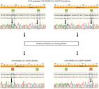
          To the Editor: The 2019 novel coronavirus (SARS-CoV-2) epidemic, which was first reported in December 2019 in Wuhan, China, and has been declared a public health emergency of international concern by the World Health Organization, may progress to a pandemic associated with substantial morbidity and mortality. SARS-CoV-2 is genetically related to SARS-CoV, which caused a global epidemic with 8096 confirmed cases in more than 25 countries in 2002–2003. 1 The epidemic of SARS-CoV was successfully contained through public health interventions, including case detection and isolation. Transmission of SARS-CoV occurred mainly after days of illness 2 and was associated with modest viral loads in the respiratory tract early in the illness, with viral loads peaking approximately 10 days after symptom onset. 3 We monitored SARS-CoV-2 viral loads in upper respiratory specimens obtained from 18 patients (9 men and 9 women; median age, 59 years; range, 26 to 76) in Zhuhai, Guangdong, China, including 4 patients with secondary infections (1 of whom never had symptoms) within two family clusters (Table S1 in the Supplementary Appendix, available with the full text of this letter at NEJM.org). The patient who never had symptoms was a close contact of a patient with a known case and was therefore monitored. A total of 72 nasal swabs (sampled from the mid-turbinate and nasopharynx) (Figure 1A) and 72 throat swabs (Figure 1B) were analyzed, with 1 to 9 sequential samples obtained from each patient. Polyester flock swabs were used for all the patients. From January 7 through January 26, 2020, a total of 14 patients who had recently returned from Wuhan and had fever (≥37.3°C) received a diagnosis of Covid-19 (the illness caused by SARS-CoV-2) by means of reverse-transcriptase–polymerase-chain-reaction assay with primers and probes targeting the N and Orf1b genes of SARS-CoV-2; the assay was developed by the Chinese Center for Disease Control and Prevention. Samples were tested at the Guangdong Provincial Center for Disease Control and Prevention. Thirteen of 14 patients with imported cases had evidence of pneumonia on computed tomography (CT). None of them had visited the Huanan Seafood Wholesale Market in Wuhan within 14 days before symptom onset. Patients E, I, and P required admission to intensive care units, whereas the others had mild-to-moderate illness. Secondary infections were detected in close contacts of Patients E, I, and P. Patient E worked in Wuhan and visited his wife (Patient L), mother (Patient D), and a friend (Patient Z) in Zhuhai on January 17. Symptoms developed in Patients L and D on January 20 and January 22, respectively, with viral RNA detected in their nasal and throat swabs soon after symptom onset. Patient Z reported no clinical symptoms, but his nasal swabs (cycle threshold [Ct] values, 22 to 28) and throat swabs (Ct values, 30 to 32) tested positive on days 7, 10, and 11 after contact. A CT scan of Patient Z that was obtained on February 6 was unremarkable. Patients I and P lived in Wuhan and visited their daughter (Patient H) in Zhuhai on January 11 when their symptoms first developed. Fever developed in Patient H on January 17, with viral RNA detected in nasal and throat swabs on day 1 after symptom onset. We analyzed the viral load in nasal and throat swabs obtained from the 17 symptomatic patients in relation to day of onset of any symptoms (Figure 1C). Higher viral loads (inversely related to Ct value) were detected soon after symptom onset, with higher viral loads detected in the nose than in the throat. Our analysis suggests that the viral nucleic acid shedding pattern of patients infected with SARS-CoV-2 resembles that of patients with influenza 4 and appears different from that seen in patients infected with SARS-CoV. 3 The viral load that was detected in the asymptomatic patient was similar to that in the symptomatic patients, which suggests the transmission potential of asymptomatic or minimally symptomatic patients. These findings are in concordance with reports that transmission may occur early in the course of infection 5 and suggest that case detection and isolation may require strategies different from those required for the control of SARS-CoV. How SARS-CoV-2 viral load correlates with culturable virus needs to be determined. Identification of patients with few or no symptoms and with modest levels of detectable viral RNA in the oropharynx for at least 5 days suggests that we need better data to determine transmission dynamics and inform our screening practices.
            Bookmark
            • Record: found
            • Abstract: found
            • Article: found
            Is Open Access

            Escape from neutralizing antibodies by SARS-CoV-2 spike protein variants

            Neutralizing antibodies elicited by prior infection or vaccination are likely to be key for future protection of individuals and populations against SARS-CoV-2. Moreover, passively administered antibodies are among the most promising therapeutic and prophylactic anti-SARS-CoV-2 agents. However, the degree to which SARS-CoV-2 will adapt to evade neutralizing antibodies is unclear. Using a recombinant chimeric VSV/SARS-CoV-2 reporter virus, we show that functional SARS-CoV-2 S protein variants with mutations in the receptor-binding domain (RBD) and N-terminal domain that confer resistance to monoclonal antibodies or convalescent plasma can be readily selected. Notably, SARS-CoV-2 S variants that resist commonly elicited neutralizing antibodies are now present at low frequencies in circulating SARS-CoV-2 populations. Finally, the emergence of antibody-resistant SARS-CoV-2 variants that might limit the therapeutic usefulness of monoclonal antibodies can be mitigated by the use of antibody combinations that target distinct neutralizing epitopes.
              Bookmark
              • Record: found
              • Abstract: found
              • Article: not found

              SARS-CoV-2 Omicron variant replication in human bronchus and lung ex vivo

              The emergence of SARS-CoV-2 variants of concern with progressively increased transmissibility between humans is a threat to global public health. The Omicron variant of SARS-CoV-2 also evades immunity from natural infection or vaccines1, but it is unclear whether its exceptional transmissibility is due to immune evasion or intrinsic virological properties. Here we compared the replication competence and cellular tropism of the wild-type virus and the D614G, Alpha (B.1.1.7), Beta (B.1.351), Delta (B.1.617.2) and Omicron (B.1.1.529) variants in ex vivo explant cultures of human bronchi and lungs. We also evaluated the dependence on TMPRSS2 and cathepsins for infection. We show that Omicron replicates faster than all other SARS-CoV-2 variants studied in the bronchi but less efficiently in the lung parenchyma. All variants of concern have similar cellular tropism compared to the wild type. Omicron is more dependent on cathepsins than the other variants of concern tested, suggesting that the Omicron variant enters cells through a different route compared with the other variants. The lower replication competence of Omicron in the human lungs may explain the reduced severity of Omicron that is now being reported in epidemiological studies, although determinants of severity are multifactorial. These findings provide important biological correlates to previous epidemiological observations.
                Bookmark

                Author and article information

                Contributors
                stephane.nizet@glock.at
                Journal
                Sci Rep
                Sci Rep
                Scientific Reports
                Nature Publishing Group UK (London )
                2045-2322
                22 March 2023
                22 March 2023
                2023
                : 13
                : 4673
                Affiliations
                [1 ]Glock Health, Science and Research GmbH, Hausfeldstrasse 17, 2232 Deutsch-Wagram, Austria
                [2 ]GRID grid.11598.34, ISNI 0000 0000 8988 2476, Diagnostic and Research Institute of Pathology, , Medical University Graz, ; Neue Stiftingtalstrasse 6, 8010 Graz, Austria
                [3 ]Wernberg, Austria
                Article
                31744
                10.1038/s41598-023-31744-z
                10031168
                36949092
                9caa6bb5-9be9-4257-9c8b-4d058386f74e
                © The Author(s) 2023

                Open Access This article is licensed under a Creative Commons Attribution 4.0 International License, which permits use, sharing, adaptation, distribution and reproduction in any medium or format, as long as you give appropriate credit to the original author(s) and the source, provide a link to the Creative Commons licence, and indicate if changes were made. The images or other third party material in this article are included in the article's Creative Commons licence, unless indicated otherwise in a credit line to the material. If material is not included in the article's Creative Commons licence and your intended use is not permitted by statutory regulation or exceeds the permitted use, you will need to obtain permission directly from the copyright holder. To view a copy of this licence, visit http://creativecommons.org/licenses/by/4.0/.

                History
                : 25 October 2022
                : 16 March 2023
                Categories
                Article
                Custom metadata
                © The Author(s) 2023

                Uncategorized
                sars-cov-2,viral epidemiology,viral transmission
                Uncategorized
                sars-cov-2, viral epidemiology, viral transmission

                Comments

                Comment on this article