29
views
0
recommends
+1 Recommend
0 collections
    0
    shares
      • Record: found
      • Abstract: found
      • Article: found
      Is Open Access

      Streptococcus gallolyticus subsp. gallolyticus promotes colorectal tumor development

      research-article

      Read this article at

          There is no author summary for this article yet. Authors can add summaries to their articles on ScienceOpen to make them more accessible to a non-specialist audience.

          Abstract

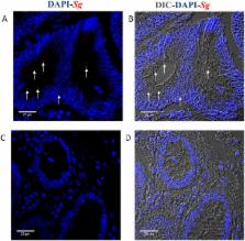
          Streptococcus gallolyticus subsp. gallolyticus ( Sg) has long been known to have a strong association with colorectal cancer (CRC). This knowledge has important clinical implications, and yet little is known about the role of Sg in the development of CRC. Here we demonstrate that Sg promotes human colon cancer cell proliferation in a manner that depends on cell context, bacterial growth phase and direct contact between bacteria and colon cancer cells. In addition, we observed increased level of β-catenin, c-Myc and PCNA in colon cancer cells following incubation with Sg. Knockdown or inhibition of β-catenin abolished the effect of Sg. Furthermore, mice administered with Sg had significantly more tumors, higher tumor burden and dysplasia grade, and increased cell proliferation and β-catenin staining in colonic crypts compared to mice receiving control bacteria. Finally, we showed that Sg is present in the majority of CRC patients and is preferentially associated with tumor compared to normal tissues obtained from CRC patients. These results taken together establish for the first time a tumor-promoting role of Sg that involves specific bacterial and host factors and have important clinical implications.

          Author summary

          Colorectal cancer (CRC) is a leading cause of cancer-related death. The recognition that microbial agents can contribute to the development of CRC raises hope for improving CRC diagnosis and treatment by incorporating both microbial and patient characteristics into clinical strategies. S. gallolyticus subsp. gallolyticus ( Sg) has been implicated in CRC for decades. Patients with Sg infections display a much higher risk of having CRC compared to the general population. Despite this, the precise role of Sg in the development of CRC— i. e., whether this organism plays an active role in the development of tumor or its presence is merely a consequence of the tumor environment being favorable for its colonization of the colon—was unknown. Here using in vitro cell cultures and mouse models of CRC, we demonstrate that Sg actively promotes colon cancer cell proliferation and tumor growth, suggesting that it is not an innocent “passenger”. These results represent a major step forward in understanding the relationship between Sg and CRC. This combined with the prevalence of Sg in CRC patients highlight Sg as being both clinically relevant and functionally important for CRC.

          Related collections

          Most cited references57

          • Record: found
          • Abstract: found
          • Article: not found

          Identification of c-MYC as a target of the APC pathway.

          The adenomatous polyposis coli gene (APC) is a tumor suppressor gene that is inactivated in most colorectal cancers. Mutations of APC cause aberrant accumulation of beta-catenin, which then binds T cell factor-4 (Tcf-4), causing increased transcriptional activation of unknown genes. Here, the c-MYC oncogene is identified as a target gene in this signaling pathway. Expression of c-MYC was shown to be repressed by wild-type APC and activated by beta-catenin, and these effects were mediated through Tcf-4 binding sites in the c-MYC promoter. These results provide a molecular framework for understanding the previously enigmatic overexpression of c-MYC in colorectal cancers.
            Bookmark
            • Record: found
            • Abstract: found
            • Article: not found

            Wnt/beta-catenin signaling in cancer stemness and malignant behavior.

            Stem cells are defined by their intrinsic capacity to self-renew and differentiate. Cancer stem cells retain both these features but have lost homeostatic mechanisms which maintain normal cell numbers. The canonical Wnt/beta-catenin signaling pathway plays a central role in modulating the delicate balance between stemness and differentiation in several adult stem cell niches such as the hair follicles in the skin, the mammary gland, and the intestinal crypt. Accordingly, constitutive Wnt signaling activation, resulting from mutations in genes encoding its downstream components, underlies tumorigenesis in these tissues. In the majority of sporadic colorectal cancer cases, the rate-limiting event is either loss of APC function or oncogenic beta-catenin mutations. However, although the presence of these initiating mutations would predict nuclear beta-catenin accumulation throughout the tumor mass, heterogeneous intracellular distributions of this key Wnt signaling molecule are observed within primary tumors and their metastases. In particular, tumor cells located at the invasive front and those migrating into the adjacent stromal tissues show nuclear beta-catenin staining. Hence, different levels of Wnt signaling activity reflect tumor heterogeneity and are likely to account for distinct cellular activities such as proliferation and epithelial-mesenchymal transitions, which prompt tumor growth and malignant behavior, respectively. Several intrinsic (cell-autonomous and/or autocrine) and extrinsic (paracrine, derived from the tumor microenvironment) factors may explain this heterogeneity of Wnt/beta-catenin signaling activity within the tumor mass.
              Bookmark
              • Record: found
              • Abstract: found
              • Article: not found

              Caught up in a Wnt storm: Wnt signaling in cancer.

              The Wnt signaling pathway, named for its most upstream ligands, the Wnts, is involved in various differentiation events during embryonic development and leads to tumor formation when aberrantly activated. Molecular studies have pinpointed activating mutations of the Wnt signaling pathway as the cause of approximately 90% of colorectal cancer (CRC), and somewhat less frequently in cancers at other sites, such as hepatocellular carcinoma (HCC). Ironically, Wnts themselves are only rarely involved in the activation of the pathway during carcinogenesis. Mutations mimicking Wnt stimulation-generally inactivating APC mutations or activating beta-catenin mutations-result in nuclear accumulation of beta-catenin which subsequently complexes with T-cell factor/lymphoid enhancing factor (TCF/LEF) transcription factors to activate gene transcription. Recent data identifying target genes has revealed a genetic program regulated by beta-catenin/TCF controlling the transcription of a suite of genes promoting cellular proliferation and repressing differentiation during embryogenesis, carcinogenesis, and in the post-embryonic regulation of cell positioning in the intestinal crypts. This review considers the spectra of tumors arising from active Wnt signaling and attempts to place perspective on recent data that begin to elucidate the mechanisms prompting uncontrolled cell growth following induction of Wnt signaling.
                Bookmark

                Author and article information

                Contributors
                Role: Editor
                Journal
                PLoS Pathog
                PLoS Pathog
                plos
                plospath
                PLoS Pathogens
                Public Library of Science (San Francisco, CA USA )
                1553-7366
                1553-7374
                13 July 2017
                July 2017
                : 13
                : 7
                : e1006440
                Affiliations
                [1 ] Center for Infectious and Inflammatory Diseases, Institute of Biosciences and Technology, Texas A&M Health Science Center, Houston, Texas, United States of America
                [2 ] Department of Pathology, Baylor College of Medicine, Houston, Texas, United States of America
                [3 ] Department of Gastrointestinal Medical Oncology, the University of Texas M. D. Anderson Cancer Center, Houston, Texas, United States of America
                [4 ] Epidemiology, the University of Texas M. D. Anderson Cancer Center, Houston, Texas, United States of America
                [5 ] Department of Internal Medicine, University of Texas Health Science Center, Houston, Texas, United States of America
                [6 ] Department of Microbiology and Microbial Genetics, University of Texas Health Science Center, Houston, Texas, United States of America
                [7 ] Harbin Institute of Technology, Harbin, China
                [8 ] Program in Integrative Nutrition and Complex Diseases, Texas A&M University, College Station, Texas, United States of America
                [9 ] Center for Epigenetics and Disease Prevention, Institute of Biosciences and Technology, Texas A&M Health Science Center, Houston, Texas, United States of America
                [10 ] Center for Translational Cancer Research, Institute of Biosciences and Technology, Texas A&M Health Science Center, Houston, Texas, United States of America
                [11 ] Department of Microbial Pathogenesis and Immunology, College of Medicine, Texas A&M Health Science Center, College Station, Texas, United States of America
                University of California, San Francisco, UNITED STATES
                Author notes

                The authors have declared that no competing interests exist.

                • Conceptualization: RK JLH YX CM.

                • Funding acquisition: YX.

                • Investigation: RK JLH DS.

                • Methodology: RK JLH FH YX EC RSC WMD RHD CM TB.

                • Project administration: RK YX.

                • Resources: RK JLH YX JD SK MMM BEM.

                • Supervision: YX RK.

                • Visualization: RK YX.

                • Writing – original draft: RK JLH YX.

                • Writing – review & editing: RK YX JD SK MMM BEM YL EC RSC WMD RHD CM.

                Author information
                http://orcid.org/0000-0001-9456-1682
                http://orcid.org/0000-0001-9647-3416
                http://orcid.org/0000-0002-2473-846X
                http://orcid.org/0000-0002-1747-6799
                Article
                PPATHOGENS-D-16-02488
                10.1371/journal.ppat.1006440
                5509344
                28704539
                7639f442-4785-4cc9-95e8-de1614ab0e47
                © 2017 Kumar et al

                This is an open access article distributed under the terms of the Creative Commons Attribution License, which permits unrestricted use, distribution, and reproduction in any medium, provided the original author and source are credited.

                History
                : 9 November 2016
                : 31 May 2017
                Page count
                Figures: 9, Tables: 2, Pages: 31
                Funding
                Funded by: Hamill Foundation
                Award Recipient :
                The study was supported by funds from the Hamill Foundation and Texas A&M Health Science Center. JD is supported in part by a grant from The University of Texas MD Anderson Cancer Center Duncan Family Institute for Cancer Prevention and Risk Assessment.Characterized Cell Line Core Facility, University of Texas MD Anderson Cancer Center, Houston, TX is funded by NCI # CA016672. The funders had no role in study design, data collection and analysis, decision to publish, or preparation of the manuscript.
                Categories
                Research Article
                Medicine and Health Sciences
                Oncology
                Cancers and Neoplasms
                Colorectal Cancer
                Biology and Life Sciences
                Anatomy
                Digestive System
                Gastrointestinal Tract
                Colon
                Medicine and Health Sciences
                Anatomy
                Digestive System
                Gastrointestinal Tract
                Colon
                Research and Analysis Methods
                Experimental Organism Systems
                Model Organisms
                Mouse Models
                Research and Analysis Methods
                Model Organisms
                Mouse Models
                Research and Analysis Methods
                Experimental Organism Systems
                Animal Models
                Mouse Models
                Biology and Life Sciences
                Cell Biology
                Cell Processes
                Cell Proliferation
                Biology and Life Sciences
                Organisms
                Bacteria
                Lactococcus
                Lactococcus Lactis
                Biology and Life Sciences
                Microbiology
                Medical Microbiology
                Microbial Pathogens
                Bacterial Pathogens
                Lactococcus
                Lactococcus Lactis
                Medicine and Health Sciences
                Pathology and Laboratory Medicine
                Pathogens
                Microbial Pathogens
                Bacterial Pathogens
                Lactococcus
                Lactococcus Lactis
                Research and analysis methods
                Biological cultures
                Cell lines
                HT29 cells
                Medicine and Health Sciences
                Oncology
                Cancer Treatment
                Research and analysis methods
                Biological cultures
                Cell lines
                SW480 cells
                Custom metadata
                All relevant data are within the paper and its Supporting Information files.

                Infectious disease & Microbiology
                Infectious disease & Microbiology

                Comments

                Comment on this article