1
views
0
recommends
+1 Recommend
0 collections
    0
    shares
      • Record: found
      • Abstract: found
      • Article: found
      Is Open Access

      Masson’s tumor involving the hand: A case report

      case-report

      Read this article at

          There is no author summary for this article yet. Authors can add summaries to their articles on ScienceOpen to make them more accessible to a non-specialist audience.

          Highlights

          • Masson’s tumor or IPEH is a rare benign tumor composed of reactive endothelial cells in a thrombus.

          • Angiosarcoma may mimick Masson’s tumor, thus ruling it out is imperative.

          • Surgical resection conveys excellent outcome and prognosis, with handful of recurrence cases.

          Abstract

          Introduction

          Masson’s tumor or IPEH represents a rare exuberant endothelial proliferation within a thrombus through an uncomprehended phenomenon. Being reported for the 1st time in Saudi Arabia, plastic surgeons should keep it in the list of differential diagnosis.

          Case

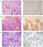
          We report a case of 17-year-old-girl who presented with a 6-month-old, painful mass on the volar side of her left 4th MCP. Radiology was inconclusive. Histopathology reported Masson’s tumor following surgical excision with good functional outcome and no recurrence.

          Discussion

          Comprising 2%–4% of overall skin vascular tumor and with no identifying clinical or radiological feature, IPEH poses as a diagnostic challenge. The literature reports similar tumors in the hand with different locations and presentations. Surgical excision remains the cornerstone of management, yet the role of radiotherapy remains undefined. Incomplete excision may result in recurrence, which requires a consensus on the extend of marginal excision. Rare cases of recurrence were reported. Histopathology is the only reliable method of diagnosis.

          Conclusion

          We present the 1st reported case of IPEH involving the hand in Saudi Arabia. Plastic surgeons should be aware of such rare conditions and be able to differentiate them from relatively identical yet more sinister tumors, especially angiosarcoma.

          Related collections

          Most cited references18

          • Record: found
          • Abstract: found
          • Article: not found

          Intravascular papillary endothelial hyperplasia. A clinicopathologic study of 91 cases.

          Ninety-one cases of intravascular papillary endothelial hyperplasia were studied clinically and histologically. This peculiar benign process, occasionally resembling hemangiosarcoma, was subgrouped in the following categories: a pure form that occurs within a dilated vascular space (30 cases), a mixed form that appears as a focal change in a hemangioma (55 cases), a third form (six cases) that belongs to neither of the first two. In the pure form, the lesions were most frequently situated in the subcutis of fingers (14 cases), of the head and neck (seven cases), and in the region between the elbows and hands (six cases). In the mixed form, half of the accompanying hemangiomas were intramuscular in no particular predilective sites. Papillary proliferation of endothelial cells was commonly found to be closely associated with thrombotic material and seemed to be an unusual feature of a thrombus undergoing organization.
            Bookmark
            • Record: found
            • Abstract: found
            • Article: not found

            Intravascular papillary endothelial hyperplasia.

            Papillary endothelial hyperplasia is a peculiar benign intravascular process that bears a remarkable resemblance to a hemangiosarcoma. In 44 cases of this lesion studied from the files of the Armed Forces Institute of Pathology, the process manifested as a small tumor-like lesion that occurred most frequently in the subcutis of the fingers (14 cases), the head and neck region (ten), and the trunk (seven). Microscopically, the tuft-like or papillary proliferation of endothelial cells was nearly always intimately associated with a thrombus and seemed to represent a peculiar variant of an organizing process. Features that aided in recognition and differential diagnosis from a hemangio-sarcoma included the intraluminal location of the lesion, the absence of tissue necrosis, and the intimate association of the proliferated tuft-like structures with thrombotic material. Follow-up information obtained in 31 cases indicated a benign clinical course despite the sarcoma-like microscopic appearance of this condition.
              Bookmark
              • Record: found
              • Abstract: found
              • Article: found
              Is Open Access

              Clinicopathological features and prognostic factors in angiosarcoma: A retrospective analysis of 200 patients from a single Chinese medical institute

              Angiosarcoma is a rare soft tissue sarcoma, and the data about its clinicopathological features and prognostic factors are limited. The purpose of the present study was to report a large series of angiosarcoma at a single institution. Clinical data from 200 cases of angiosarcoma from the Shanghai Cancer Center (Shanghai, China) between March 2006 and March 2014 were retrospectively analyzed. The study population included 97 males and 103 females with ages between 4 and 91 years (median, 53 years). According to the tumor location, 200 cases were divided into 4 groups: i) Tumors involving the head and neck; ii) breast; iii) viscera (including internal organs and bone); and iv) soft tissue (including trunk and extremities). Of the 113 patients with follow-up data, 46 patients succumbed to the disease with a median interval of 10 months. Tumor recurrence/metastasis was identified in 66 patients with a median interval of 4 months. The disease-free survival (DFS) rate at 5-years was 19.3% and the overall survival (OS) rate at 5-years was 40.8%. Site of tumor origin, size (≥5 cm) and histological differentiation influenced DFS (P=0.032, 0.038 and <0.001, respectively), and OS (P<0.001, 0.008 and <0.001, respectively) rates. Age (<65 years) and multimodal treatment correlated with improved OS (P=0.003 and <0.001, respectively). Tumor differentiation and treatment modality were identified to be independent determinants of OS (P<0.001 and 0.038, respectively). Tumor recurrence/metastasis was an independent predictor of DFS (P<0.001). The prognosis of angiosarcoma is poor and the mortality rate is high. The site of tumor origin, size, histological differentiation, age, treatment modality and tumor recurrence/metastasis are all significant prognostic factors. In the present study, multimodal treatment may improve the clinical outcome of patients with angiosarcoma.
                Bookmark

                Author and article information

                Contributors
                Journal
                Int J Surg Case Rep
                Int J Surg Case Rep
                International Journal of Surgery Case Reports
                Elsevier
                2210-2612
                08 May 2020
                2020
                08 May 2020
                : 70
                : 223-226
                Affiliations
                [a ]King Faisal Specialist Hospital and Research Center, Department of Surgery, Plastic and Reconstructive Surgery Section, Riyadh, Saudi Arabia
                [b ]Alfaisal University, College of Medicine, Riyadh, Saudi Arabia
                Author notes
                [* ]Corresponding author at: PO Box: 3354, 11211, Saudi Arabia. Almarghoubm@ 123456gmail.com
                Article
                S2210-2612(20)30262-5
                10.1016/j.ijscr.2020.04.069
                7229427
                32422583
                d0e24e9e-b261-4d41-96b7-76aa9388f983
                © 2020 The Author(s)

                This is an open access article under the CC BY license (http://creativecommons.org/licenses/by/4.0/).

                History
                : 29 February 2020
                : 3 April 2020
                : 19 April 2020
                Categories
                Article

                masson’s tumor,intravascular papillary endothelial hyperplasia,ipeh,hand,case report

                Comments

                Comment on this article