29
views
0
recommends
+1 Recommend
0 collections
    0
    shares
      • Record: found
      • Abstract: found
      • Article: found
      Is Open Access

      Intramedullary Spinal Cysticercosis: A Case Report and Review of Literature

      case-report

      Read this article at

      Bookmark
          There is no author summary for this article yet. Authors can add summaries to their articles on ScienceOpen to make them more accessible to a non-specialist audience.

          Abstract

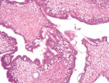
          To report a case of spinal intramedullary cysticercosis in thoracic spine. A 47-year old man living in Korea referred to our hospital with both feet tingling sensation for about a year. Laboratory evaluations, including serologic tests were not helpful. Magnetic resonance imaging revealed a 1.7 cm intramedullary mass at T10-11 level, which believed to be a tumor instead, rather than a cysticercosis preoperatively. Successful operation was done with a histopathological result confirmed it as cysticercosis. Even though the prevalence of intramedullary spinal cysticercosis is extremely rare, and radiologic exams mimic other common tumors like ependymoma or astrocytoma, the disease should be considered as differential diagnosis.

          Related collections

          Most cited references15

          • Record: found
          • Abstract: found
          • Article: not found

          Surgical considerations in treatment of intraventricular cysticercosis. An analysis of 45 cases.

          Infestations of the human brain with the larval stage of Taenia solium, once an infrequent diagnosis in the United States, is now a more frequently encountered clinical entity especially in population centers with high immigrant flux. During a recent 5-year period 45 cases of intraventricular cysticerosis have been evaluated and treated. Modes of involvement included isolated cyst formation, ependymitis, or combinations of both. Evidence of associated parenchymatous involvement was present in 20% of cases. Sites of infestation included the lateral ventricle (five cases), third ventricle (12 cases), Sylvian aqueduct (four cases), and fourth ventricle (24 cases). Mean post-therapy follow-up periods for this series exceed 36 months. This experience indicates that direct excision is the treatment of choice for ventricular cystic lesions, but that management, operative planning, and expectations should be influenced by considerations of: 1) the potential for acute clinical deterioration (38%); 2) the potential for cyst migration; 3) attendant ependymitis, defined by computerized tomography or verified at surgery; 4) the potential for increase in cyst volume with local mass effect; 5) selection and institution of corridors of surgical access that establish alternative routes of cerebrospinal fluid flow; and 6) the possibility of cyst excision by a stereotaxic endoscopic procedure.
            Bookmark
            • Record: found
            • Abstract: found
            • Article: not found

            Treatment of intramedullary spinal cysticercosis: report of 2 cases and review of literature.

            NCC, Taenia solium involvement in the central nervous system, is one of the most common parasitic diseases. Spinal IMC is one of the rarest forms of NCC. We report 2 cases (an 8-year-old child and a 35-year-old woman) of spinal IMC. Both cysts were dorsal in location. MRI was diagnostic in both cases. One case was treated surgically because of acute presentation. The other cyst healed with treatment with albendazole. Both patients had good clinical outcomes. With medical management, IMC may take 1 year to resolve on MRI.
              Bookmark
              • Record: found
              • Abstract: found
              • Article: not found

              Intramedullary cysticercosis cured with drug treatment. A case report.

              A report of a patient with cervical intramedullary cysticercosis is presented. To report the first case of intramedullary. cysticercosis cured with drug management only, which supports the hypothesis that management with anthelmintics might be successfully used in this form of cysticercosis. Intramedullary cysticercosis is a rare manifestation of neurocysticercosis. Every treated patient reported to date has undergone surgery, frequently necessary for diagnosis. Since the anthelmintics praziquantel and albendazole were shown to be effective in parenchymal brain cysticercosis, these drugs have been considered potentially useful in patients with intramedullary cysticercosis. Nevertheless, no case yet had been reported to be cared with only medical therapy. The treatment of a patient who suffered multiple cysticercal reinfestations of the nervous system is presented. The patient received prolonged treatment with albendazole because of superimposed cerebral reinfestations. During this treatment, she suffered acute paraparesis, and cervical magnetic resonance imaging showed cyst-like lesions with linear gadolinium enhancement and perilesional edema, indicative of dying cysticerci and inflammatory host reaction. Dexamethasone was added, and progressive neurologic improvement followed with complete resolution of intramedullary lesions. A preoperative diagnostic suspicion of cysticercosis is important in patients with intramedullary cystic lesions because specific drug treatment is available. Treatment with anthelmintics, particularly albendazole, should be considered in patients with intramedullary cysticercosis before surgery. Corticosteroids may be added to the therapeutic regimen because this may reduce the perilesional edema and prevent neurologic deterioration during the course of anthelmintic treatment.
                Bookmark

                Author and article information

                Journal
                Korean J Spine
                Korean J Spine
                KJS
                Korean Journal of Spine
                The Korean Spinal Neurosurgery Society
                1738-2262
                2093-6729
                June 2014
                30 June 2014
                : 11
                : 2
                : 81-84
                Affiliations
                [1 ]Department of Neurological Surgery, Asan Medical Center, College of Medicine, University of Ulsan College of Medicine, Seoul, Korea.
                [2 ]Department of Pathology, Asan Medical Center, College of Medicine, University of Ulsan College of Medicine, Seoul, Korea.
                Author notes
                Corresponding Author: Seung Chul Rhim, MD, PhD. Department of Neurological Surgery, Asan Medical Center, University of Ulsan College of Medicine, 86 Asanbyeongwon-gil, Songpa-gu, Seoul 138-736, Korea. Tel: +82-2-3010-3555, Fax: +82-2-476-6738, aussie84@ 123456naver.com
                Article
                10.14245/kjs.2014.11.2.81
                4124922
                25110489
                d288b2b9-0444-4028-9e54-6b8c95eca602
                Copyright © 2014 The Korean Spinal Neurosurgery Society

                This is an Open Access article distributed under the terms of the Creative Commons Attribution Non-Commercial License ( http://creativecommons.org/licenses/by-nc/3.0/) which permits unrestricted non-commercial use, distribution, and reproduction in any medium, provided the original work is properly cited.

                History
                : 12 April 2014
                : 02 May 2014
                : 09 May 2014
                Categories
                Case Report

                cysticercosis,intramedullary,thoracic spine
                cysticercosis, intramedullary, thoracic spine

                Comments

                Comment on this article