23
views
0
recommends
+1 Recommend
0 collections
    0
    shares
      • Record: found
      • Abstract: found
      • Article: not found

      Plasmodium falciparum Histones Induce Endothelial Proinflammatory Response and Barrier Dysfunction

      research-article

      Read this article at

      ScienceOpenPublisherPMC
          There is no author summary for this article yet. Authors can add summaries to their articles on ScienceOpen to make them more accessible to a non-specialist audience.

          Abstract

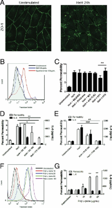
          Plasmodium falciparum is a protozoan parasite of human erythrocytes that causes the most severe form of malaria. Severe P. falciparum infection is associated with endothelial activation and permeability, which are important determinants of the outcome of the infection. How endothelial cells become activated is not fully understood, particularly with regard to the effects of parasite subcomponents. We demonstrated that P. falciparum histones extracted from merozoites (HeH) directly stimulated the production of IL-8 and other inflammatory mediators by primary human dermal microvascular endothelial cells through a signaling pathway that involves Src family kinases and p38 MAPK. The stimulatory effect of HeH and recombinant P. falciparum H3 (PfH3) was abrogated by histone-specific antibodies. The release of nuclear contents on rupture of infected erythrocytes was captured by live cell imaging and confirmed by detecting nucleosomes in the supernatants of parasite cultures. HeH and recombinant parasite histones also induced endothelial permeability through a charge-dependent mechanism that resulted in disruption of junctional protein expression and cell death. Recombinant human activated protein C cleaved HeH and PfH3 and abrogated their proinflammatory effects. Circulating nucleosomes of both human and parasite origin were detected in the plasma of patients with falciparum malaria and correlated positively with disease severity. These results support a pathogenic role for both host- and pathogen-derived histones in P. falciparum-caused malaria.

          Related collections

          Most cited references51

          • Record: found
          • Abstract: found
          • Article: not found

          Malaria hemozoin is immunologically inert but radically enhances innate responses by presenting malaria DNA to Toll-like receptor 9.

          Hemozoin (HZ) is an insoluble crystal formed in the food vacuole of malaria parasites. HZ has been reported to induce inflammation by directly engaging Toll-like receptor (TLR) 9, an endosomal receptor. "Synthetic" HZ (beta-hematin), typically generated from partially purified extracts of bovine hemin, is structurally identical to natural HZ. When HPLC-purified hemin was used to synthesize the crystal, beta-hematin had no inflammatory activity. In contrast, natural HZ from Plasmodium falciparum cultures was a potent TLR9 inducer. Natural HZ bound recombinant TLR9 ectodomain, but not TLR2. Both TLR9 stimulation and TLR9 binding of HZ were abolished by nuclease treatment. PCR analysis demonstrated that natural HZ is coated with malarial but not human DNA. Purified malarial DNA activated TLR9 but only when DNA was targeted directly to the endosome with a transfection reagent. Stimulatory quantities of natural HZ contain <1 microg of malarial DNA; its potency in activating immune responses was even greater than transfecting malarial DNA. Thus, although the malarial genome is extremely AT-rich, its DNA is highly proinflammatory, with the potential to induce cytokinemia and fever during disease. However, its activity depends on being bound to HZ, which we propose amplifies the biological responses to malaria DNA by targeting it to a TLR9(+) intracellular compartment.
            Bookmark
            • Record: found
            • Abstract: found
            • Article: not found

            Toll-like receptor 9 mediates innate immune activation by the malaria pigment hemozoin

            Malaria parasites within red blood cells digest host hemoglobin into a hydrophobic heme polymer, known as hemozoin (HZ), which is subsequently released into the blood stream and then captured by and concentrated in the reticulo-endothelial system. Accumulating evidence suggests that HZ is immunologically active, but the molecular mechanism(s) through which HZ modulates the innate immune system has not been elucidated. This work demonstrates that HZ purified from Plasmodium falciparum is a novel non-DNA ligand for Toll-like receptor (TLR)9. HZ activated innate immune responses in vivo and in vitro, resulting in the production of cytokines, chemokines, and up-regulation of costimulatory molecules. Such responses were severely impaired in TLR9−/− and myeloid differentiation factor 88 (MyD88)−/−, but not in TLR2, TLR4, TLR7, or Toll/interleukin 1 receptor domain–containing adaptor-inducing interferon β−/− mice. Synthetic HZ, which is free of the other contaminants, also activated innate immune responses in vivo in a TLR9-dependent manner. Chloroquine (CQ), an antimalarial drug, abrogated HZ-induced cytokine production. These data suggest that TLR9-mediated, MyD88-dependent, and CQ-sensitive innate immune activation by HZ may play an important role in malaria parasite–host interactions.
              Bookmark
              • Record: found
              • Abstract: found
              • Article: not found

              Induction of proinflammatory responses in macrophages by the glycosylphosphatidylinositols of Plasmodium falciparum: cell signaling receptors, glycosylphosphatidylinositol (GPI) structural requirement, and regulation of GPI activity.

              The glycosylphosphatidylinositol (GPI) anchors of Plasmodium falciparum have been proposed to be the major factors that contribute to malaria pathogenesis through their ability to induce proinflammatory responses. In this study we identified the receptors for P. falciparum GPI-induced cell signaling that leads to proinflammatory responses and studied the GPI structure-activity relationship. The data show that GPI signaling is mediated mainly through recognition by TLR2 and to a lesser extent by TLR4. The activity of sn-2-lyso-GPIs is comparable with that of the intact GPIs, whereas the activity of Man(3)-GPIs is about 80% that of the intact GPIs. The GPIs with three (intact GPIs and Man(3)-GPIs) and two fatty acids (sn-2-lyso-GPIs) appear to differ considerably in the requirement of the auxiliary receptor, TLR1 or TLR6, for recognition by TLR2. The former are preferentially recognized by TLR2/TLR1, whereas the latter are favored by TLR2/TLR6. However, the signaling pathways initiated by all three GPI types are similar, involving the MyD88-dependent activation of extracellular signal-regulated kinase, c-Jun N-terminal kinase, and p38 and NF-kappaB-signaling pathways. The signaling molecules of these pathways differentially contribute to the production of various cytokines and nitric oxide (Zhu, J., Krishnegowda, G., and Gowda, D. C. (2004) J. Biol. Chem. 280, 8617-8627). Our data also show that GPIs are degraded by the macrophage surface phospholipases predominantly into inactive species, indicating that the host can regulate GPI activity at least in part by this mechanism. These results imply that macrophage surface phospholipases play important roles in the GPI-induced innate immune responses and malaria pathogenesis.
                Bookmark

                Author and article information

                Journal
                Am J Pathol
                Am. J. Pathol
                The American Journal of Pathology
                American Society for Investigative Pathology
                0002-9440
                1525-2191
                March 2012
                March 2012
                : 180
                : 3
                : 1028-1039
                Affiliations
                [* ]Department of Microbiology Immunology & Infectious Diseases, University of Calgary, Alberta, Canada
                []Department of Biochemistry and Molecular Biology, Pennsylvania State University College of Medicine, Hershey, Pennsylvania
                []Temple Autoimmunity Center, Department of Microbiology and Immunology, Temple University School of Medicine, Philadelphia, Pennsylvania
                [§ ]Department of Entomology, Pennsylvania State University, University Park, Pennsylvania
                []Oxford University Clinical Research Unit, Hospital for Tropical Diseases, Ho Chi Minh City, Vietnam
                Author notes
                [* ]Address reprint requests to May Ho, M.D., Department of Microbiology and Infectious Diseases, 3330 Hospital Dr. NW, Calgary, AB, Canada T2N 4N1 mho@ 123456ucalgary.ca
                Article
                AJPA754
                10.1016/j.ajpath.2011.11.037
                3448071
                22260922
                dd7d2b80-046d-4d46-8200-da77e026ac57
                © 2012 American Society for Investigative Pathology. Published by Elsevier Inc. All rights reserved.

                This document may be redistributed and reused, subject to certain conditions.

                History
                : 4 November 2011
                Categories
                Regular Article
                Immunopathology and Infectious Diseases

                Pathology
                Pathology

                Comments

                Comment on this article