15
views
0
recommends
+1 Recommend
0 collections
    0
    shares
      • Record: found
      • Abstract: found
      • Article: found
      Is Open Access

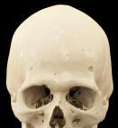
      Syphilitic osteomyelitis in a patient with headache and lytic lesions

      other
      , MD, , MD, , MD, , MD MEd
      CMAJ : Canadian Medical Association Journal
      CMA Impact Inc.

      Read this article at

          There is no author summary for this article yet. Authors can add summaries to their articles on ScienceOpen to make them more accessible to a non-specialist audience.

          Related collections

          Most cited references11

          • Record: found
          • Abstract: found
          • Article: not found

          Bone involvement in secondary syphilis: a case report and systematic review of the literature.

          Bone involvement is an unusual manifestation of secondary syphilis, but little information is available in the English-language literature. We carried out a systematic review of the English-language literature from 1964 to 2013, describing cases of secondary syphilis with bone involvement. We also describe a case of secondary syphilis with multiple osteolytic lesions, mimicking metastatic cancer or myeloma, which was included in an analysis of 37 eligible cases of secondary syphilis with bone involvement. Of these 37 patients, 28 (76%) patients were male, and the median age was 32 years (range, 12-64 years). Eleven (30%) patients had human immunodeficiency virus (HIV) infection with a median CD4 lymphocyte count of 343 cells/mm (range, 130-689 cells/mm). The diagnosis of early syphilis was suspected based on mucocutaneous findings in 28 (76%) cases. In the remaining 9 (24%) cases, high titers of nontreponemal serologic tests were the only evidence of early syphilis. The median venereal disease research laboratory (VDRL) titer was 1:64 (range, 1:8-1:320), and median rapid plasma reagin (RPR) titer was 1:64 (range, 1:16-1:512). The bones most often affected were long bones of the limbs (n = 22) and skull (n = 21). The bone lesions were multifocal in 27 (73%) cases and osteolytic in 19 (51%) cases. The treatment of syphilitic bone lesions was medical only in most patients, and prognosis was favorable with high-dose penicillin therapy. Clinical features and outcome between HIV-uninfected and HIV-infected patients were not different. Knowledge of this rare entity may lead to early diagnosis and appropriate management.
            Bookmark
            • Record: found
            • Abstract: found
            • Article: not found

            Osteomyelitis of the skull in early-acquired syphilis: evaluation by MR imaging and CT.

            We present an unusual case of acquired secondary syphilis manifesting as osteomyelitis of the skull in a patient with a history of human immunodeficiency virus infection, evaluated by CT, volumetric CT reconstructions, and MR imaging.
              Bookmark
              • Record: found
              • Abstract: found
              • Article: found
              Is Open Access

              Calvarial osteomyelitis in secondary syphilis: evaluation by MRI and CT, including cinematic rendering

              This is a case of a 22-year-old, HIV-negative, male patient with asymptomatic syphilitic osteomyelitis of the skull in the context of secondary syphilis. The diagnosis was made based on serology as well as CT and MRI scans. CT volumetric data was post-processed with cinematic rendering, which is a novel algorithm that allows for a photorealistic visualization of the lesions. Imaging and follow-up scans after treatment confirmed the diagnosis without the need to perform invasive procedures such as a biopsy.
                Bookmark

                Author and article information

                Journal
                CMAJ
                CMAJ
                9711805
                CMAJ : Canadian Medical Association Journal
                CMA Impact Inc.
                0820-3946
                1488-2329
                4 March 2024
                04 March 2024
                : 196
                : 8
                : E260-E264
                Affiliations
                Department of Medicine (Bogler, Said, Sharma), and Division of Infectious Diseases (Said, Sharma), Department of Medicine, and Department of Medical Imaging (Mathur), University of Toronto; Division of Infectious Diseases (Sharma), St. Michael’s Hospital, Unity Health Toronto Department of Medical Imaging (Mathur), St. Michael’s Hospital, Unity Health Toronto; Medical Imaging Office of Research & Innovation (Mathur), Li Ka Shing Knowledge Institute, Toronto, Ont.
                Author notes
                Correspondence to: Malika Sharma, malika.sharma@ 123456utoronto.ca
                Article
                196e260
                10.1503/cmaj.230666
                10911860
                38438150
                16280278-6c4f-48a5-b23d-c1404a12066b
                © 2024 CMA Impact Inc. or its licensors

                This is an Open Access article distributed in accordance with the terms of the Creative Commons Attribution (CC BY-NC-ND 4.0) licence, which permits use, distribution and reproduction in any medium, provided that the original publication is properly cited, the use is noncommercial (i.e., research or educational use), and no modifications or adaptations are made. See: https://creativecommons.org/licenses/by-nc-nd/4.0/

                History
                Categories
                Practice
                Cases

                Comments

                Comment on this article