15
views
0
recommends
+1 Recommend
0 collections
    0
    shares
      • Record: found
      • Abstract: found
      • Article: found
      Is Open Access

      Assessment of Third Molar Impaction Pattern and Associated Clinical Symptoms in a Central Anatolian Turkish Population

      research-article

      Read this article at

          There is no author summary for this article yet. Authors can add summaries to their articles on ScienceOpen to make them more accessible to a non-specialist audience.

          Abstract

          Objectives

          The purpose of this study was to assess the pattern of third molar impaction and associated symptoms in a Central Anatolian Turkish population.

          Material and Methods

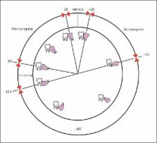
          A total of 2,133 impacted third molar teeth of 705 panoramic radiographs were reviewed. The positions of impacted third molar teeth on the panoramic radiographs were documented according to the classifications of Pell and Gregory and of Winter. The presence of related symptoms including pain, pericoronitis, lymphadenopathy and trismus was noted for every patient. Distributions of obtained values were compared using the Pearson χ 2 test. Nonparametric values were analyzed using the Mann-Whitney U test and Kruskal-Wallis test.

          Results

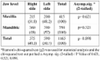
          The mean age of the subjects was 30.58 ± 11.98 years (range: 19-73); in a review of the 2,133 impacted third molar teeth, the most common angulation of impaction in both maxillaries was vertical (1,177; 55%). Level B impaction was the most common in the maxilla (425/1,037; 39%), while level C impaction was the most common in the mandible (635/1,096; 61%). Pain (272/705; 39%) and pericoronitis (188/705; 27%) were found to be the most common complications of impaction. Among 705 patients (335 males, 370 females), pericoronitis was more prevalent in males (101; 30%) and usually related to lower third molars (236; 22%). The retromolar space was significantly smaller in females (p < 0.05). Moreover, there was a significant difference in retromolar space for the area of jaw (maxillary: 11.3 mm; mandibular: 14.2 mm) and impaction level (A: 14.7 mm; B: 11.1 mm; C: 10.3 mm; p < 0.05).

          Conclusion

          The pattern of third molar impaction in a Central Anatolian Turkish population was characterized by a high prevalence rate of level C impaction with vertical position. Pain and pericoronitis were the most common symptoms usually associated with level A impaction and vertical position.

          Related collections

          Most cited references30

          • Record: found
          • Abstract: found
          • Article: not found

          Extraction of impacted mandibular third molars: postoperative complications and their risk factors.

          The purpose of this prospective study was to evaluate the incidence of various complications, including alveolitis, infection and paresthesia of the inferior alveolar nerve, in association with removal of impacted mandibular third molars. The relation between these 3 complications and several clinical variables (age, sex, degree of impaction, surgical difficulty and use of oral contraceptives) was also examined. Data were collected prospectively for all patients who underwent extraction of an impacted third molar in a single private dental practice over a 12-month period. A variety of data were collected for each patient, including age, sex, medical status at the time of the procedure and type of procedure performed. Patients were contacted at 2 days and 4 weeks after surgery to establish the occurrence of complications, and those with complications were treated; those with paresthesia were followed for at least 24 months. A total of 550 impacted mandibular third molars were extracted from 327 patients (136 males and 191 females). The complication rate was 6.9%, consisting of 20 cases of alveolitis, 12 cases of infection and 6 cases of paresthesia of the inferior alveolar nerve. Of the 6 neurosensory deficits, 3 resolved and 3 were permanent. The risk factors associated with permanent neurosensory deficit were female sex, Pell and Gregory IC or IIC classification of impaction, and age greater than 24 years. The risk of postoperative alveolitis and infection was also greater among women. There was no significant relation between the use of oral contraceptives and alveolitis. Surgical removal of impacted mandibular third molars should be carried out well before the age of 24 years, especially for female patients. Older patients are at greater risk of postoperative complications and permanent sequelae. A surgeon's lack of experience could also be a major factor in the development of postoperative complications.
            Bookmark
            • Record: found
            • Abstract: found
            • Article: found
            Is Open Access

            Incidence of impacted mandibular and maxillary third molars: a radiographic study in a Southeast Iran population

            Objectives: The aim of this study is to evaluate the position of impacted third molars based on the classifications of Pell & Gregory and Winter in a sample of Iranian patients. Study design: In this retrospective study, up to 1020 orthopantomograms (OPG) of the patients who were referred to the radiology clinics from October 2007 to January 2011 were evaluated. Data including the age, gender, the angulation type, width and depth of impaction were evaluated by statistical tests. Results: Among 1020 patients, 380(27.3%) were male and 640(62.7%) were female with the sex ratio was 1:1.7. Of the 1020 OPGs, 585 cases showed at least one impacted third molar, with significant difference between males (205; 35.1%) and females (380; 64.9%) (P = 0.0311). Data analysis showed that impacted third molars were 1.9 times more likely to occur in the mandible than in the maxilla (P =0.000). The most common angulation of impaction in the mandible was mesioangular impaction (48.3%) and the most common angulation of impaction in the maxilla was the vertical (45.3%). Impaction in the level IIA was the most common in both maxilla and mandible. There was no significant diffe-rence between the right and left sides in both the maxilla and the mandible. Conclusion: The pattern of third molar impaction in the southeast region of Iran is characterized by a high prevalence of impaction, especially in the mandible. Female more than male have teeth impaction. The most common angulation was the mesioangular in the mandible, and the vertical angulation in the maxilla. The most common level of impaction was the A and there was no any significant difference between the right and left sides in both jaws. Key words:Third molar, impaction, incidence, Iran.
              Bookmark
              • Record: found
              • Abstract: found
              • Article: not found

              Reliability of panoramic radiography in evaluating the topographic relationship between the mandibular canal and impacted third molars.

              The authors conducted a study to evaluate the predictive value of five radiographic markers on the panoramic radiograph, or PR, to point out the relationship between the mandibular canal and the impacted third molar. The authors evaluated the accuracy of the radiographic markers by comparing the PR with an axial computed tomographic, or CT, scan. They identified a sample of 73 third molars that showed a close relationship between the tooth roots and the mandibular canal on the PR, and then classified them on the basis of five radiographic markers. They also detected contact between the third molar and the mandibular canal on the CT scan. The distribution of the five radiographic markers was as follows: 37 teeth exhibited increased radiolucency, 13 exhibited superimposition, 14 exhibited interruption of the radiopaque border, 14 exhibited narrowing of the canal and seven exhibited diversion of the canal. In 11 cases, two or more markers were recognizable. The predictive values of a positive test result were as follows: increased radiolucency, 73 percent; superimposition, 38.5 percent; interruption of the radiopaque border, 71.4 percent; narrowing, 78.6 percent; and diversion, 100 percent. The authors detected contact in all of the cases that exhibited two or more markers. Increased radiolucency, narrowing and interruption of the radiopaque border, as well as the concomitant presence of two or more radiographic markers, on the PR were highly predictive of contact between the third molar and the mandibular canal. An axial CT scan probably is indicated in such cases. The results of this study may lead to some guidelines for oral surgeons evaluating whether to obtain an axial CT scan for further investigation after examining an impacted mandibular third molar via PR.
                Bookmark

                Author and article information

                Journal
                Med Princ Pract
                Med Princ Pract
                MPP
                Medical Principles and Practice
                S. Karger AG (Allschwilerstrasse 10, P.O. Box · Postfach · Case postale, CH–4009, Basel, Switzerland · Schweiz · Suisse, Phone: +41 61 306 11 11, Fax: +41 61 306 12 34, karger@karger.ch )
                1011-7571
                1423-0151
                February 2016
                13 November 2015
                13 November 2015
                : 25
                : 2
                : 169-175
                Affiliations
                [1] aDepartment of Oral and Maxillofacial Radiology, Faculty of Dentistry, Kırıkkale University, Kırıkkale, Turkey
                [2] bDepartment of Statistics, Faculty of Science and Letters, Kırıkkale University, Kırıkkale, Turkey
                Author notes
                *Dr. Mehmet Zahit Adìsen, Department of Oral and Maxillofacial Radiology, Faculty of Dentistry, Kırıkkale University, TR—71450 Kırıkkale (Turkey), E-Mail m_zahit@ 123456hotmail.com
                Article
                mpp-0025-0169
                10.1159/000442416
                5588352
                26566129
                343677f6-9dfd-4714-9f17-09aad39532a0
                Copyright © 2015 by S. Karger AG, Basel

                This is an Open Access article licensed under the terms of the Creative Commons Attribution-NonCommercial 3.0 Unported license (CC BY-NC) (www.karger.com/OA-license), applicable to the online version of the article only. Distribution permitted for non-commercial purposes only.

                History
                : 24 March 2015
                : 12 November 2015
                Page count
                Figures: 3, Tables: 5, References: 30, Pages: 7
                Categories
                Original Paper

                third molar impaction,winter's classification,pain,pericoronitis,turkish population

                Comments

                Comment on this article