3
views
0
recommends
+1 Recommend
0 collections
    0
    shares
      • Record: found
      • Abstract: found
      • Article: found
      Is Open Access

      Interobserver variability in gross tumor volume contouring in non-spine bone metastases

      research-article

      Read this article at

          There is no author summary for this article yet. Authors can add summaries to their articles on ScienceOpen to make them more accessible to a non-specialist audience.

          Abstract

          Background and Aim:

          The optimal imaging test for gross tumor volume (GTV) delineation in non-spine bone metastases has not been defined. The use of stereotactic body radiotherapy (SBRT) requires accurate target delineation. Magnetic resonance imaging (MRI) and/or 18fludesoxyglucose positron emission tomography (18FDG-PET) allow for better visualization of the extent of bone metastases and optimizes the accuracy of tumor delineation for stereotactic radiotherapy compared to computed tomography (CT) alone. We evaluated the interobserver agreement in GTV of non-spine bone metastases in a single center and compared MRI and/or 18FDG-PET and CT in GTV delineation.

          Methods:

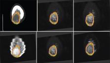
          Anonymous CT and MRI and/or 18FDG-PET obtained from 10 non-spine bone metastases were analyzed by six radiation oncologists at our center. Images acquired by CT and MRI and/or 18FDG-PET were used to delineate 10 GTVs of non-spine bone metastases in the pelvis, extremities, and skull. The cases showed different characteristics: blastic and lytic metastases, and different primary cancers (lung, breast, prostate, rectum, urothelial, and biliary). In both CT and MRI and/or 18FDG-PET, the GTV volumes were compared. The index of agreement was evaluated according to Landis and Koch protocol.

          Results:

          The GTV volume as defined on MRI was in all cases larger or at least as large as the GTV volume on CT ( P=0.25). The median GTV volume on MRI was 3.15 cc (0.027-70.64 cc) compared to 2.8 cc on CT (0.075-77.95 cc). Interobserver variance and standard deviation were lower in CT than MRI (576.3 vs. 722.2 and 24.0 vs. 26.9, respectively). The level of agreement was fair (kappa=0.36) between CT and MRI. The median GTV volume on 18FDG-PET in five patients was 5.8 cc (0.46-64.17 cc), compared to 4.1 cc on CT (0.99-54.2 cc) ( P=0.236). Interobserver variance and standard deviation in CT, MRI, and 18FDG-PET were 576.3 versus 722.2 versus 730.5 and 24 versus 26.9 versus 27.0, respectively. The level of agreement was slight (kappa=0.08) between CT and 18FDG-PET.

          Conclusions:

          Interobserver variance in non-spine bone metastases was equal when MRI and PET were compared to CT. CT was associated with the lowest variance and standard deviation. Compared to CT GTVs, the GTVs rendered from MRI images had fair agreement, while the GTVs rendered from 18FDG-PET had only slight agreement.

          Relevance for Patients:

          The delimitation of the treatment volume in non-spine bone metastases with SBRT is important for the results determining its efficacy. It is therefore essential to know the variability and to manage it to achieve the highest quality of treatment.

          Related collections

          Most cited references18

          • Record: found
          • Abstract: found
          • Article: not found

          Tumor response to radiotherapy regulated by endothelial cell apoptosis.

          About 50% of cancer patients receive radiation therapy. Here we investigated the hypothesis that tumor response to radiation is determined not only by tumor cell phenotype but also by microvascular sensitivity. MCA/129 fibrosarcomas and B16F1 melanomas grown in apoptosis-resistant acid sphingomyelinase (asmase)-deficient or Bax-deficient mice displayed markedly reduced baseline microvascular endothelial apoptosis and grew 200 to 400% faster than tumors on wild-type microvasculature. Thus, endothelial apoptosis is a homeostatic factor regulating angiogenesis-dependent tumor growth. Moreover, these tumors exhibited reduced endothelial apoptosis upon irradiation and, unlike tumors in wild-type mice, they were resistant to single-dose radiation up to 20 grays (Gy). These studies indicate that microvascular damage regulates tumor cell response to radiation at the clinically relevant dose range.
            Bookmark
            • Record: found
            • Abstract: not found
            • Article: not found

            American Society for Therapeutic Radiology and Oncology (ASTRO) and American College of Radiology (ACR) practice guideline for the performance of stereotactic body radiation therapy.

              Bookmark
              • Record: found
              • Abstract: found
              • Article: not found

              Engaging the vascular component of the tumor response.

              Recent research has shed new light on the critical role of tissue microvasculature in regulating the tumor response to radiation and drugs. In this issue of Cancer Cell, Moeller et al.(2005) demonstrate that HIF-1 activation during the course of fractionated radiotherapy initiates pleiotropic adaptive responses in both tumor cells and the microvascular network, radiosensitizing tumor cells but concomitantly conferring tumor radioresistance due to protection of the microvascular endothelium. HIF-1 thus serves as a legitimate target for differential modulation of tissue response to radiation.
                Bookmark

                Author and article information

                Journal
                J Clin Transl Res
                J Clin Transl Res
                Whioce Publishing Pte. Ltd.
                Journal of Clinical and Translational Research
                Whioce Publishing Pte. Ltd.
                2382-6533
                2424-810X
                29 December 2022
                22 October 2022
                : 8
                : 6
                : 465-469
                Affiliations
                [A1] 1Department of Radiation Oncology, Ramón y Cajal University Hospital, IRYCIS, Madrid, Spain
                [A2] 2Department of Radiology, Ramón y Cajal University Hospital, IRYCIS, Madrid, Spain
                [A3] 3Department of Nuclear Medicine, Ramón y Cajal University Hospital, IRYCIS, Madrid, Spain
                Author notes
                [* ] Corresponding author: Carolina de la Pinta Department of Radiation Oncology, Ramón y Cajal University Hospital, IRYCIS, Alcalá University, Madrid, Spain. E-mail: cdelapinta88@ 123456gmail.com
                Article
                jctres.08.202206.003
                9706312
                36452000
                4e13cb88-c4fc-43f9-a724-263a34527ec1
                Copyright: © 2022 Author(s).

                This is an Open-Access article distributed under the terms of the Creative Commons Attribution-Noncommercial License, permitting all noncommercial use, distribution, and reproduction in any medium, provided the original work is properly cited.

                History
                : 22 June 2022
                : 20 August 2022
                : 14 September 2022
                Categories
                Original Article

                stereotactic body radiotherapy,non-spine bone metastases,contouring,gross tumor volume,variability

                Comments

                Comment on this article