6
views
0
recommends
+1 Recommend
0 collections
    0
    shares
      • Record: found
      • Abstract: found
      • Article: found
      Is Open Access

      Axillary arch (of Langer): A large‐scale dissection and simulation study based on unembalmed cadavers of body donors

      research-article

      Read this article at

          There is no author summary for this article yet. Authors can add summaries to their articles on ScienceOpen to make them more accessible to a non-specialist audience.

          Abstract

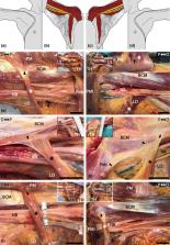
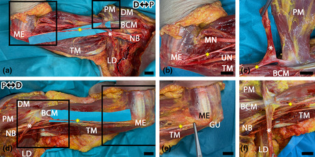
          Connective or muscular tissue crossing the axilla is named axillary arch (of Langer). It is known to complicate axillary surgery and to compress nerves and vessels transiting from the axilla to the arm. Our study aims at systematically researching the frequency, insertions, tissue composition and dimension of axillary arches in a large cohort of individuals with regard to gender and bilaterality. In addition, it aims at evaluating the ability of axillary arches to cause compression of the axillary neurovascular bundle. Four hundred axillae from 200 unembalmed and previously unharmed cadavers were investigated by careful anatomical dissection. Identified axillary arches were examined for tissue composition and insertion. Length, width and thickness were measured. The relation of the axillary arch and the neurovascular axillary bundle was recorded after passive arm movements. Twenty‐seven axillae of 18 cadavers featured axillary arches. Macroscopically, 15 solely comprised muscular tissue, six connective tissue and six both. Their average length was 79.56 mm, width 7.44 mm and thickness 2.30 mm. One to three distinct insertions were observed. After passive abduction and external rotation of the arm, 17 arches (63%) touched the neurovascular axillary bundle. According to our results, 9% of the Central European population feature an axillary arch. Approximately 50% of it bilaterally. A total of 40.74% of the arches have a thickness of 3 mm or more and 63% bear the potential of touching or compressing the neuromuscular axillary bundle upon arm movement.

          Abstract

          Nine percent of the Central European population has an axillary arch. Approximately half of these are bilateral. A total of 40.74% of the arches have a thickness of 3 mm or more, and 63% can touch or compress the neuromuscular axillary bundle during arm movements.

          Related collections

          Most cited references51

          • Record: found
          • Abstract: found
          • Article: not found

          The Incidence of Thoracic Outlet Syndrome

          Thoracic outlet syndrome (TOS) refers to a constellation of compressive problems that occur at the thoracic outlet. TOS has been recognized since the 19th century, and the "modern" era of treatment, especially for neurogenic TOS, dates from at least the 1970s. Despite this, however, the incidence and prevalence of these syndromes are almost completely unknown. To attempt to answer this fundamental question, we established a prospective database of all patients who presented to our clinic with a diagnosis of potential TOS, as described below.
            Bookmark
            • Record: found
            • Abstract: found
            • Article: not found

            Abnormal muscles that may affect axillary lymphadenectomy: surgical anatomy.

            Purpose The present study aimed at summarizing and presenting the anomalous muscles that a surgeon might encounter during axillary lymphadenectomy (AL). Methods For this purpose, both the anatomical and surgical literature was reviewed and an anatomical study on 107 cadavers was carried out. Furthermore, based on the anatomical features of the anomalous muscles that came up during our study and taking into consideration the landmarks of the AL, we further analyzed the complications that may arise from each of these muscles, along with their preoperative and intraoperative recognition and management. Results The literature review revealed that there are three supernumerary muscles that may affect the AL, namely the Langer's axillary arch, the pectoralis quartus and the chondroepitrochlearis muscles, as well as the aplasia of the lower part of the pectoralis major muscle. Eight out of the 107 (7.48%) cadavers that we dissected had such an abnormal muscle in the axilla. Specifically, the axillary arch was found unilaterally in five cadavers (4.67%) and the pectoralis quartus muscle was present unilaterally in three cadavers (2.8%). One cadaver had both an axillary arch and a pectoralis quartus muscle in the right side. The abdominal and almost the whole sternocostal portion of the pectoralis major as well the pectoralis minor muscle were absent in one cadaver (0.93%). The chondroepitrochlearis muscle was not found in any of the cadavers that we dissected. Conclusions The present study offers the necessary preoperative knowledge for recognizing these muscles during AL, avoiding thus the complications that may arise from them.
              Bookmark
              • Record: found
              • Abstract: found
              • Article: not found

              Contribution to the anatomical nomenclature concerning upper limb anatomy.

              The aim of this article is to revise and extend the existing sections of Terminologia Anatomica dealing with the upper limb structures, which nomenclature belongs to its most neglected and not developing parts, and to justify the use of the proposed anatomical terms in the clinical practice, research, and education.
                Bookmark

                Author and article information

                Contributors
                paata.pruidze@meduniwien.ac.at
                Journal
                J Anat
                J Anat
                10.1111/(ISSN)1469-7580
                JOA
                Journal of Anatomy
                John Wiley and Sons Inc. (Hoboken )
                0021-8782
                1469-7580
                15 November 2023
                March 2024
                15 November 2023
                : 244
                : 3 ( doiID: 10.1111/joa.v244.3 )
                : 448-457
                Affiliations
                [ 1 ] Division of Anatomy Medical University of Vienna Vienna Austria
                [ 2 ] Department of Neurosurgery, Neuromed Campus Kepler University Hospital Linz Austria
                Author notes
                [*] [* ] Correspondence

                Paata Pruidze, Waehringer str. 13, Vienna 1090, Austria.

                Email: paata.pruidze@ 123456meduniwien.ac.at

                Author information
                https://orcid.org/0000-0001-7618-9979
                https://orcid.org/0000-0003-2283-4439
                Article
                JOA13976 JANAT-2023-0332.R2
                10.1111/joa.13976
                10862185
                37965841
                6072884a-81bb-4ad2-8dc2-aaf8d965363e
                © 2023 The Authors. Journal of Anatomy published by John Wiley & Sons Ltd on behalf of Anatomical Society.

                This is an open access article under the terms of the http://creativecommons.org/licenses/by-nc-nd/4.0/ License, which permits use and distribution in any medium, provided the original work is properly cited, the use is non‐commercial and no modifications or adaptations are made.

                History
                : 30 October 2023
                : 14 July 2023
                : 02 November 2023
                Page count
                Figures: 5, Tables: 1, Pages: 10, Words: 5625
                Categories
                Original Article
                Original Articles
                Custom metadata
                2.0
                March 2024
                Converter:WILEY_ML3GV2_TO_JATSPMC version:6.3.6 mode:remove_FC converted:13.02.2024

                Anatomy & Physiology
                axilla,axillopectoral muscle,chondroepitrochlearis muscle,thoracic outlet syndrome,variation

                Comments

                Comment on this article