7
views
0
recommends
+1 Recommend
1 collections
    0
    shares
      • Record: found
      • Abstract: found
      • Article: not found

      Lethal pediatric cerebral vasculitis triggered by Severe Acute Respiratory Syndrome Coronavirus 2: a case report

      brief-report

      Read this article at

      ScienceOpenPublisherPMC
          There is no author summary for this article yet. Authors can add summaries to their articles on ScienceOpen to make them more accessible to a non-specialist audience.

          Abstract

          Objectives

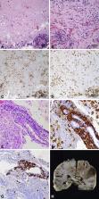
          We report the clinical, radiological, laboratory, and neuropathological findings in support of the first diagnosis of lethal, small-vessel cerebral vasculitis triggered by severe acute respiratory syndrome coronavirus 2 (SARS-CoV-2) in a pediatric patient.

          Patient description

          A previously healthy, 8-year-old Hispanic female presented with subacute onset left-sided weakness two weeks after a mild febrile illness. SARS-CoV-2 nasopharyngeal swab was positive. Magnetic resonance imaging (MRI) revealed an enhancing right frontal lobe lesion with significant vasogenic edema. Two brain biopsies of the lesion showed perivascular and intraluminal lymphohistiocytic inflammatory infiltrate consistent with vasculitis. Despite extensive treatment with immunomodulatory therapies targeting primary angiitis of the central nervous system, she experienced neurologic decline and died 93 days after presentation. SARS-CoV-2 testing revealed positive serum IgG and positive cerebrospinal fluid IgM. Comprehensive infectious, rheumatologic, hematologic/oncologic, and genetic evaluation did not identify an alternative etiology. Post-mortem brain autopsy remained consistent with vasculitis.

          Discussion

          This is the first pediatric presentation to suggest SARS-CoV-2 can lead to a fatal, post-infectious, inflammatory small-vessel cerebral vasculitis. Our case is unique in including supportive cerebrospinal fluid and post-mortem tissue analysis. While most children recover well from neurologic complications of SARS-CoV-2, we emphasize the potential mortality in a child with no risk factors for severe disease.

          Related collections

          Most cited references13

          • Record: found
          • Abstract: found
          • Article: not found

          Neurologic Involvement in Children and Adolescents Hospitalized in the United States for COVID-19 or Multisystem Inflammatory Syndrome

          Coronavirus disease 2019 (COVID-19) affects the nervous system in adult patients. The spectrum of neurologic involvement in children and adolescents is unclear.
            Bookmark
            • Record: found
            • Abstract: found
            • Article: not found

            Neuroimaging manifestations in children with SARS-CoV-2 infection: a multinational, multicentre collaborative study

            Background The CNS manifestations of COVID-19 in children have primarily been described in case reports, which limit the ability to appreciate the full spectrum of the disease in paediatric patients. We aimed to identify enough cases that could be evaluated in aggregate to better understand the neuroimaging manifestations of COVID-19 in the paediatric population. Methods An international call for cases of children with encephalopathy related to severe acute respiratory syndrome coronavirus 2 (SARS-CoV-2) infection and abnormal neuroimaging findings was made. Clinical history and associated plasma and cerebrospinal fluid data were requested. These data were reviewed by a central neuroradiology panel, a child neurologist, and a paediatric infectious diseases expert. The children were categorised on the basis of their time of probable exposure to SARS-CoV-2. In addition, cases were excluded when a direct link to SARS-CoV-2 infection could not be established or an established alternate diagnostic cause could be hypothesised. The accepted referral centre imaging data, from ten countries, were remotely reviewed by a central panel of five paediatric neuroradiologists and a consensus opinion obtained on the imaging findings. Findings 38 children with neurological disease related to SARS-CoV-2 infection were identified from France (n=13), the UK (n=8), the USA (n=5), Brazil (n=4), Argentina (n=4), India (n=2), Peru (n=1), and Saudi Arabia (n=1). Recurring patterns of disease were identified, with neuroimaging abnormalities ranging from mild to severe. The most common imaging patterns were postinfectious immune-mediated acute disseminated encephalomyelitis-like changes of the brain (16 patients), myelitis (eight patients), and neural enhancement (13 patients). Cranial nerve enhancement could occur in the absence of corresponding neurological symptoms. Splenial lesions (seven patients) and myositis (four patients) were predominantly observed in children with multisystem inflammatory syndrome. Cerebrovascular complications in children were less common than in adults. Significant pre-existing conditions were absent and most children had favourable outcomes. However, fatal atypical CNS co-infections developed in four previously healthy children infected with SARS-CoV-2. Interpretation Acute-phase and delayed-phase SARS-CoV-2-related CNS abnormalities are seen in children. Recurring patterns of disease and atypical neuroimaging manifestations can be found and should be recognised being as potentially due to SARS-CoV-2 infection as an underlying aetiological factor. Studies of paediatric specific cohorts are needed to better understand the effects of SARS-CoV-2 infection on the CNS at presentation and on long-term follow-up in children. Funding American Society of Pediatric Neuroradiology, University of Manchester (Manchester, UK). Video Abstract Neuroimaging manifestations in children with SARS-CoV-2 infection
              Bookmark
              • Record: found
              • Abstract: found
              • Article: found
              Is Open Access

              Classification of Extrapulmonary Manifestations Due to Mycoplasma pneumoniae Infection on the Basis of Possible Pathogenesis

              The list of extrapulmonary manifestations due to Mycoplasma pneumoniae infection can be classified according to the following three possible mechanisms derived from the established biological activity of M. pneumoniae; (1) a direct type in which the bacterium is present at the site of inflammation and local inflammatory cytokines induced by the bacterium play an important role (2) an indirect type in which the bacterium is not present at the site of inflammation and immune modulations, such as autoimmunity or formation of immune complexes, play an important role, and (3) a vascular occlusion type in which obstruction of blood flow induced either directly or indirectly by the bacterium plays an important role. Recent studies concerning extrapulmonary manifestations have prompted the author to upgrade the list, including cardiac and aortic thrombi as cardiovascular manifestations; erythema nodosum, cutaneous leukocytoclastic vasculitis, and subcorneal pustular dermatosis as dermatological manifestations; acute cerebellar ataxia, opsoclonus-myoclonus syndrome, and thalamic necrosis as neurological manifestations; pulmonary embolism as a respiratory system manifestation; and renal artery embolism as a urogenital tract manifestation. Continuing nosological confusion on M. pneumoniae–induced mucositis (without skin lesions), which may be called M. pneumoniae-associated mucositis or M. pneumoniae-induced rash and mucositis separately from Stevens-Johnson syndrome, is argued in the dermatological manifestations. Serological methods are recommended for diagnosis because pneumonia or respiratory symptoms are often minimal or even absent in extrapulmonary manifestations due to M. pneumoniae infection. Concomitant use of immunomodulators, such as corticosteroids or immunoglobulins with antibiotics effective against M. pneumoniae, can be considered as treatment modalities for most severe cases, such as encephalitis. Further studies would be necessary to construct a comprehensive therapeutic strategy, covering microbiology (antibiotics), immunology (immunomodulators), and hematology (anticoagulants). The possible influence of the emergence of macrolide-resistant M. pneumoniae on extrapulmonary manifestations, which can be considered of limited clinical threat in Japan where the resistant rate has currently decreased, is discussed on the basis of unique biological characteristics of M. pneumoniae, the smallest self-replicating organism.
                Bookmark

                Author and article information

                Journal
                Pediatr Neurol
                Pediatr Neurol
                Pediatric Neurology
                Elsevier Inc.
                0887-8994
                1873-5150
                12 November 2021
                12 November 2021
                Affiliations
                [1 ]University of Cincinnati College of Medicine, Department of Pediatrics
                [2 ]Cincinnati Children’s Hospital Medical Center, Division of Child Neurology
                [3 ]Cincinnati Children’s Hospital Medical Center, Division of Pathology
                [4 ]State University of New York, Upstate Medical University, Department of Pathology
                [5 ]Cincinnati Children’s Hospital Medical Center, Division of Radiology
                [6 ]Cincinnati Children’s Hospital Medical Center, Division of Infectious Disease
                [7 ]Cincinnati Children’s Hospital Medical Center, Division of Rheumatology
                [8 ]Cincinnati Children’s Hospital Medical Center, Department of Pediatrics
                [9 ]Cincinnati Children’s Hospital Medical Center, Division of Neurosurgery
                Author notes
                [] Corresponding Author: Marissa Vawter-Lee 3333 Burnet Avenue, MLC 2015, Cincinnati, Ohio, United States of America, 45229 Phone: 513-636-4222
                Article
                S0887-8994(21)00248-4
                10.1016/j.pediatrneurol.2021.11.003
                8585961
                2b9dbbbe-12fa-4a85-8a82-ba4e695084f2
                © 2021 Elsevier Inc. All rights reserved.

                Since January 2020 Elsevier has created a COVID-19 resource centre with free information in English and Mandarin on the novel coronavirus COVID-19. The COVID-19 resource centre is hosted on Elsevier Connect, the company's public news and information website. Elsevier hereby grants permission to make all its COVID-19-related research that is available on the COVID-19 resource centre - including this research content - immediately available in PubMed Central and other publicly funded repositories, such as the WHO COVID database with rights for unrestricted research re-use and analyses in any form or by any means with acknowledgement of the original source. These permissions are granted for free by Elsevier for as long as the COVID-19 resource centre remains active.

                History
                : 25 October 2021
                : 2 November 2021
                : 6 November 2021
                Categories
                Short Communication

                Pediatrics
                sars-cov-2,covid,vasculitis,pediatrics
                Pediatrics
                sars-cov-2, covid, vasculitis, pediatrics

                Comments

                Comment on this article