5
views
0
recommends
+1 Recommend
0 collections
    0
    shares
      • Record: found
      • Abstract: found
      • Article: found
      Is Open Access

      Programable Active Fixator System for Systematic In Vivo Investigation of Bone Healing Processes

      research-article

      Read this article at

          There is no author summary for this article yet. Authors can add summaries to their articles on ScienceOpen to make them more accessible to a non-specialist audience.

          Abstract

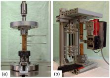
          This manuscript introduces a programable active bone fixator system that enables systematic investigation of bone healing processes in a sheep animal model. In contrast to previous systems, this solution combines the ability to precisely control the mechanical conditions acting within a fracture with continuous monitoring of the healing progression and autonomous operation of the system throughout the experiment. The active fixator system was implemented on a double osteotomy model that shields the experimental fracture from the influence of the animal’s functional loading. A force sensor was integrated into the fixator to continuously measure stiffness of the repair tissue as an indicator for healing progression. A dedicated control unit was developed that allows programing of different loading protocols which are later executed autonomously by the active fixator. To verify the feasibility of the system, it was implanted in two sheep with different loading protocols, mimicking immediate and delayed weight-bearing, respectively. The implanted devices operated according to the programmed protocols and delivered seamless data over the whole course of the experiment. The in vivo trial confirmed the feasibility of the system. Hence, it can be applied in further preclinical studies to better understand the influence of mechanical conditions on fracture healing.

          Related collections

          Most cited references44

          • Record: found
          • Abstract: not found
          • Article: not found

          Fracture healing: The diamond concept

            Bookmark
            • Record: found
            • Abstract: found
            • Article: not found

            The influence of induced micromovement upon the healing of experimental tibial fractures.

            Although it has been well established that fracture healing is influenced by the mechanical environment, the optimal parameters have not yet been established. In two groups of sheep an experimental tibial diaphysial fracture was created, and stabilised using external skeletal fixation. In one group rigid fixation was maintained throughout fracture healing; in the other group controlled axial micromovement, with a loading regime known to be osteogenic in intact bones, was applied for a short period daily. A significant improvement in healing was associated with the application of controlled micromovement. Data from these experiments provide the basis for improving the conditions for fracture healing and may assist in the prevention of delayed union.
              Bookmark
              • Record: found
              • Abstract: found
              • Article: not found

              Shear movement at the fracture site delays healing in a diaphyseal fracture model.

              This study tested the hypothesis that interfragmentary axial movement of transverse diaphyseal osteotomies would result in improved fracture healing compared to interfragmentary shear movement. Ten skeletally mature merino sheep underwent a middiaphyseal osteotomy of the right tibia, stabilized by external fixation with an interfragmentary gap of 3 mm. A custom made external fixator allowed either pure axial (n=5) or pure shear movement (n=5) of 1.5 mm amplitude during locomotion by the animals. The movement of the osteotomy gap was monitored weekly in two sheep by an extensometer temporarily attached to the fixator. After 8 weeks the sheep were killed, and healing of the osteotomies was evaluated by radiography, biomechanical testing, and undecalcified histology. Shear movement considerably delayed the healing of diaphyseal osteotomies. Bridging of the osteotomy fragments occurred in all osteotomies in the axial group (100%), while in the shear group only three osteotomies (60%) were partially bridged. Peripheral callus formation in the shear group was reduced by 36% compared to the axial group (p<0.05). In the axial group bone formation was considerably larger at the peripheral callus and in between the osteotomy gaps but not in the intramedullary area. The larger peripheral callus and excess in bone tissue at the level of the gap resulted in a more than three times larger mechanical rigidity for the axial than for the shear group (p<0.05). In summary, fixation that allows excessive shear movement significantly delayed the healing of diaphyseal osteotomies compared to healing under axial movement of the same magnitude.
                Bookmark

                Author and article information

                Journal
                Sensors (Basel)
                Sensors (Basel)
                sensors
                Sensors (Basel, Switzerland)
                MDPI
                1424-8220
                22 December 2020
                January 2021
                : 21
                : 1
                : 17
                Affiliations
                [1 ]AO Research Institute Davos, Clavadelerstrasse 8, 7270 Davos, Switzerland; manuela.ernst@ 123456aofoundation.org (M.E.); stephan.zeiter@ 123456aofoundation.org (S.Z.); markus.windolf@ 123456aofoundation.org (M.W.)
                [2 ]Bulgarian Academy of Sciences, Institute of Metal Science ‘Acad. A. Balevski’, Shipchenski prohod 67, 1574 Sofia, Bulgaria; ljudmil.d@ 123456ims.bas.bg
                [3 ]Orthopaedic Research Institute of Queensland, 7 Turner Street, Townsville, QLD 4812, Australia; constantin.dlaska@ 123456gmx.at
                [4 ]Institute of Health and Biomedical Innovation, Queensland University of Technology, George Street 2, Brisbane City, QLD 4000, Australia; d.epari@ 123456qut.edu.au
                Author notes
                [* ]Correspondence: jan.barcik@ 123456aofoundation.org ; Tel.: +41-81-414-2592
                Author information
                https://orcid.org/0000-0003-3730-5134
                https://orcid.org/0000-0002-8155-4202
                https://orcid.org/0000-0001-8077-6914
                Article
                sensors-21-00017
                10.3390/s21010017
                7792812
                33375087
                8517e738-2317-4b29-917f-b17d818c1ad4
                © 2020 by the authors.

                Licensee MDPI, Basel, Switzerland. This article is an open access article distributed under the terms and conditions of the Creative Commons Attribution (CC BY) license ( http://creativecommons.org/licenses/by/4.0/).

                History
                : 30 September 2020
                : 19 December 2020
                Categories
                Article

                Biomedical engineering
                motorized fixation,mechanobiology of fracture healing,healing monitoring,rehabilitation,surgery

                Comments

                Comment on this article