6
views
0
recommends
+1 Recommend
0 collections
    0
    shares
      • Record: found
      • Abstract: found
      • Article: found
      Is Open Access

      Wearable Photoplethysmography for Cardiovascular Monitoring

      research-article

      Read this article at

          There is no author summary for this article yet. Authors can add summaries to their articles on ScienceOpen to make them more accessible to a non-specialist audience.

          Abstract

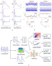
          Smart wearables provide an opportunity to monitor health in daily life and are emerging as potential tools for detecting cardiovascular disease (CVD). Wearables such as fitness bands and smartwatches routinely monitor the photoplethysmogram signal, an optical measure of the arterial pulse wave that is strongly influenced by the heart and blood vessels. In this survey, we summarize the fundamentals of wearable photoplethysmography and its analysis, identify its potential clinical applications, and outline pressing directions for future research in order to realize its full potential for tackling CVD.

          Related collections

          Most cited references291

          • Record: found
          • Abstract: found
          • Article: found
          Is Open Access

          MIMIC-III, a freely accessible critical care database

          MIMIC-III (‘Medical Information Mart for Intensive Care’) is a large, single-center database comprising information relating to patients admitted to critical care units at a large tertiary care hospital. Data includes vital signs, medications, laboratory measurements, observations and notes charted by care providers, fluid balance, procedure codes, diagnostic codes, imaging reports, hospital length of stay, survival data, and more. The database supports applications including academic and industrial research, quality improvement initiatives, and higher education coursework.
            Bookmark
            • Record: found
            • Abstract: found
            • Article: found
            Is Open Access

            An Overview of Heart Rate Variability Metrics and Norms

            Healthy biological systems exhibit complex patterns of variability that can be described by mathematical chaos. Heart rate variability (HRV) consists of changes in the time intervals between consecutive heartbeats called interbeat intervals (IBIs). A healthy heart is not a metronome. The oscillations of a healthy heart are complex and constantly changing, which allow the cardiovascular system to rapidly adjust to sudden physical and psychological challenges to homeostasis. This article briefly reviews current perspectives on the mechanisms that generate 24 h, short-term (~5 min), and ultra-short-term (<5 min) HRV, the importance of HRV, and its implications for health and performance. The authors provide an overview of widely-used HRV time-domain, frequency-domain, and non-linear metrics. Time-domain indices quantify the amount of HRV observed during monitoring periods that may range from ~2 min to 24 h. Frequency-domain values calculate the absolute or relative amount of signal energy within component bands. Non-linear measurements quantify the unpredictability and complexity of a series of IBIs. The authors survey published normative values for clinical, healthy, and optimal performance populations. They stress the importance of measurement context, including recording period length, subject age, and sex, on baseline HRV values. They caution that 24 h, short-term, and ultra-short-term normative values are not interchangeable. They encourage professionals to supplement published norms with findings from their own specialized populations. Finally, the authors provide an overview of HRV assessment strategies for clinical and optimal performance interventions.
              Bookmark
              • Record: found
              • Abstract: found
              • Article: found
              Is Open Access

              Global, Regional, and National Burden of Cardiovascular Diseases for 10 Causes, 1990 to 2015

              Background The burden of cardiovascular diseases (CVDs) remains unclear in many regions of the world. Objectives The GBD (Global Burden of Disease) 2015 study integrated data on disease incidence, prevalence, and mortality to produce consistent, up-to-date estimates for cardiovascular burden. Methods CVD mortality was estimated from vital registration and verbal autopsy data. CVD prevalence was estimated using modeling software and data from health surveys, prospective cohorts, health system administrative data, and registries. Years lived with disability (YLD) were estimated by multiplying prevalence by disability weights. Years of life lost (YLL) were estimated by multiplying age-specific CVD deaths by a reference life expectancy. A sociodemographic index (SDI) was created for each location based on income per capita, educational attainment, and fertility. Results In 2015, there were an estimated 422.7 million cases of CVD (95% uncertainty interval: 415.53 to 427.87 million cases) and 17.92 million CVD deaths (95% uncertainty interval: 17.59 to 18.28 million CVD deaths). Declines in the age-standardized CVD death rate occurred between 1990 and 2015 in all high-income and some middle-income countries. Ischemic heart disease was the leading cause of CVD health lost globally, as well as in each world region, followed by stroke. As SDI increased beyond 0.25, the highest CVD mortality shifted from women to men. CVD mortality decreased sharply for both sexes in countries with an SDI >0.75. Conclusions CVDs remain a major cause of health loss for all regions of the world. Sociodemographic change over the past 25 years has been associated with dramatic declines in CVD in regions with very high SDI, but only a gradual decrease or no change in most regions. Future updates of the GBD study can be used to guide policymakers who are focused on reducing the overall burden of noncommunicable disease and achieving specific global health targets for CVD.
                Bookmark

                Author and article information

                Contributors
                Journal
                9879073
                Proc IEEE Inst Electr Electron Eng
                Proc IEEE Inst Electr Electron Eng
                Proceedings of the IEEE. Institute of Electrical and Electronics Engineers
                0018-9219
                14 March 2022
                11 March 2022
                29 March 2022
                : 110
                : 3
                : 355-381
                Affiliations
                Research Centre for Biomedical Engineering, City, University of London, London EC1V 0HB, U.K
                Department of Public Health and Primary Care, University of Cambridge, Cambridge CB1 8RN, U.K
                Department of Electronics Engineering and the Biomedical Engineering Institute, Kaunas University of Technology, 44249 Kaunas, Lithuania
                Department of Clinical Pharmacology, King’s College London, London SE1 7EH, U.K
                Department of Biomedical Engineering, School of Biomedical Engineering and Imaging Sciences, King’s College London, King’s Health Partners, London SE1 7EU, U.K
                Author notes
                Corresponding author: Peter H. Charlton is with the Department of Biomedical Engineering, School of Biomedical Engineering and Imaging Sciences, King’s College London, King’s Health Partners, London SE1 7EU, U.K., with the Research Centre for Biomedical Engineering, City, University of London, London EC1V 0HB, U.K., and also with the Department of Public Health and Primary Care, University of Cambridge, Cambridge CB1 8RN, U.K. pc657@ 123456medschl.cam.ac.uk

                This work was supported in part by the British Heart Foundation under Grant PG/15/104/31913 and Grant FS/20/20/34626, in part by the University of Cambridge EPSRC Impact Acceleration Account, in part by the Wellcome EPSRC Centre for Medical Engineering at King’s College London under Grant WT 203148/Z/16/Z, in part by the European COST ACTION-Network for Research in Vascular Ageing under Grant CA18216 supported by the European Cooperation in Science and Technology (COST), in part by the European Regional Development Fund under Project 01.2.2-LMT-K-718-01-0030 under a grant agreement with the Research Council of Lithuania (LMTLT), and in part by the Department of Health through the National Institute for Health Research Cardiovascular MedTech Co-Operative at Guy’s and St Thomas’ NHS Foundation Trust.

                Author information
                https://orcid.org/0000-0003-3836-8655
                https://orcid.org/0000-0002-2868-485X
                https://orcid.org/0000-0002-9531-0268
                https://orcid.org/0000-0002-6879-5845
                https://orcid.org/0000-0003-4507-038X
                https://orcid.org/0000-0003-3742-5259
                Article
                EMS143850
                10.1109/JPROC.2022.3149785
                7612541
                35356509
                6d16c3ce-d453-4acf-9012-2d8d74658600

                This work is licensed under a Creative Commons Attribution 4.0 License. For more information, see https://creativecommons.org/licenses/by/4.0/

                History
                Categories
                Article

                cardiovascular (cv),photoplethysmogram (ppg),pulse wave,sensor,signal processing,smartwatch

                Comments

                Comment on this article