13
views
0
recommends
+1 Recommend
0 collections
    0
    shares
      • Record: found
      • Abstract: found
      • Article: not found

      Combined Pupilloplasty and Retropupillary Iris-Claw Intraocular Lens Implantation with DSAEK in a Patient with Traumatic Iridoplegia, Aphakia and Corneal Decompensation

      case-report

      Read this article at

      ScienceOpenPublisherPMC
          There is no author summary for this article yet. Authors can add summaries to their articles on ScienceOpen to make them more accessible to a non-specialist audience.

          Abstract

          Purpose

          To report the management of a patient with traumatic mydriasis, aphakia and corneal decompensation with a triple procedure: simultaneous pupilloplasty and retropupillary iris-claw intraocular lens (IOL) implantation combined with Descemet stripping automated endothelial keratoplasty (DSAEK).

          Results

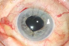
          An 88-year-old woman was referred to our Institute for consultation on her left eye. The patient had undergone surgical removal of the IOL, without re-implantation, in her left eye 10 months prior to presentation due to traumatic IOL dislocation. At the time of examination, corrected distance visual acuity was counting fingers and intraocular pressure was 10 mmHg. Slit-lamp examination revealed iridoplegia, aphakia and corneal edema. The patient underwent simultaneous pupilloplasty and retropupillary iris-claw IOL implantation combined with DSAEK. Six months postoperatively, the corneal graft was attached and clear, the iris was well reconstructed and almost round, and the iris-claw IOL was in place.

          Conclusions

          Simultaneous pupilloplasty and retropupillary iris-claw IOL implantation combined with DSAEK was shown to be a safe surgical technique in a patient with traumatic mydriasis, aphakia and corneal decompensation.

          Related collections

          Most cited references10

          • Record: found
          • Abstract: found
          • Article: not found

          Descemet-stripping automated endothelial keratoplasty.

          To evaluate the speed of visual recovery in 16 consecutive patients with corneal endothelial dysfunction who received Descemet-stripping automated endothelial keratoplasty (DSAEK). This is a retrospective study of a novel method for small-incision endothelial transplantation (DSAEK). Endothelial replacement was accomplished with Descemet stripping of the recipient and insertion of a posterior donor tissue that had been prepared with a microkeratome. Best spectacle-corrected visual acuity (BSCVA) by manifest refraction, endothelial counts, and dislocation rates were measured up to 12 months after DSAEK. Sixteen consecutive patients underwent uncomplicated DSAEK. Three patients had known optic nerve or macular disease precluding vision better than 20/200. Of the remaining 14 patients, 11 had BSCVA of 20/40 by postoperative week 12 (7 by week 6). The remaining 2 were 20/50 by weeks 6 and 12. All 14 patients were 20/40 or better at 1 year. One patient had a primary graft failure, and surgery was repeated with 20/40 BSCVA at 1 year. The dislocation rate was 25%. The average cell count between 7 and 10 months was 1714. The average pachymetry was 682. DSAEK surgery allows rapid, excellent BSCVA visual recovery. The rate of visual recovery is more rapid than usually found with penetrating keratoplasty.
            Bookmark
            • Record: found
            • Abstract: found
            • Article: not found

            Endothelial keratoplasty for Fuchs' dystrophy with cataract: complications and clinical results with the new triple procedure.

            To report the immediate postoperative complications and the 6- and 12-month clinical results in a large series of cases undergoing the new triple-procedure Descemet's stripping automated endothelial keratoplasty (DSAEK) and concurrent cataract surgery.
              Bookmark
              • Record: found
              • Abstract: not found
              • Article: not found

              Iris-claw intraocular lens for aphakia: Can location influence the final outcomes?

                Bookmark

                Author and article information

                Contributors
                konstas@med.auth.gr
                Journal
                Ophthalmol Ther
                Ophthalmol Ther
                Ophthalmology and Therapy
                Springer Healthcare (Cheshire )
                2193-8245
                2193-6528
                17 July 2019
                17 July 2019
                September 2019
                : 8
                : 3
                : 497-500
                Affiliations
                [1 ]ISNI 0000000109457005, GRID grid.4793.9, 3rd University Department of Ophthalmology, , Aristotle University, ; Thessaloniki, Greece
                [2 ]ISNI 0000 0001 2165 4204, GRID grid.9851.5, Department of Ophthalmology, , University of Lausanne, Jules-Gonin Eye Hospital, ; Fondation Asile des aveugles, Lausanne, Switzerland
                [3 ]ISNI 0000 0001 2108 7481, GRID grid.9594.1, Department of Ophthalmology, , University of Ioannina, ; Ioannina, Greece
                [4 ]ISNI 0000000109457005, GRID grid.4793.9, 1st and 3rd University Departments of Ophthalmology, , Aristotle University, ; Thessaloniki, Greece
                Article
                198
                10.1007/s40123-019-0198-2
                6692417
                31317508
                a204cff2-d76b-4610-adda-918c02d244f7
                © The Author(s) 2019
                History
                : 24 May 2019
                Categories
                Case Report
                Custom metadata
                © The Author(s) 2019

                aphakia,artisan,dmek,dsaek,endothelial keratoplasty,iridoplegia,iris-claw,ocular trauma,pupilloplasty,traumatic mydriasis

                Comments

                Comment on this article